Back
Poster Session E - Tuesday Afternoon
E0076 - A Curious Case of Recurrent Pancreatitis: A Case Report
Tuesday, October 25, 2022
3:00 PM – 5:00 PM ET
Location: Crown Ballroom

Sripriya Gonakoti, MD
Allegheny Health Network
Pittsburgh, PA
Presenting Author(s)
Sripriya Gonakoti, MD, Gianna Baker, MSc, Gursimran S. Kochhar, MD
Allegheny Health Network, Pittsburgh, PA
Introduction: Annular pancreas, a congenital anomaly characterized by incomplete rotation of the ventral pancreatic bud, causes a ring of pancreatic tissue to encircle the duodenum. It is commonly accompanied by pancreatic divisum, which occurs when the main pancreatic duct and accessory duct fail to fuse. Patients often present with recurrent pancreatitis due to partial obstruction of the pancreatic duct caused by fibrosis.
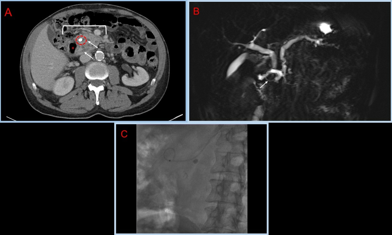
Case Description/Methods: A 72-year-old Caucasian male presented with recurrent episodes of acute pancreatitis. A CT abdomen (Image A) revealed pancreatic tissue surrounding the second portion of the duodenum consistent with an annular pancreas. A pancreatic duct stone was also noted. An MRCP (Magnetic resonance cholangiopancreatography) (Image B) was consistent with annular pancreas with persistent dilation of the main and accessory pancreatic ducts and a 6mm stone noted in the pancreatic duct. On an initial ERCP (Endoscopic retrograde cholangiopancreatography), the pancreatic duct could not be cannulated through the major papilla and minor papilla could not be identified due to inflammation. An EUS (Endoscopic ultrasound) was performed, which was suggestive of pancreas divisum. The minor papilla was cannulated, and a stent was placed in the pancreatic duct. In a subsequent ERCP (Image C), the patient had the pancreatic stone removed, with complete resolution of his symptoms.
Discussion: Typically, patients with annular pancreas present with symptoms during childhood. While infrequent, some patients remain asymptomatic until 20-50 years of age. This case demonstrates the potential to remain asymptomatic until even 72 years of age. Thus, it is important for physicians to be aware of the clinical manifestations of annular pancreas, as they could arise at any stage in life. By increasing awareness surrounding annular pancreas and management of its complications, we hope to increase the rate of early intervention.

Disclosures:
Sripriya Gonakoti, MD, Gianna Baker, MSc, Gursimran S. Kochhar, MD. E0076 - A Curious Case of Recurrent Pancreatitis: A Case Report, ACG 2022 Annual Scientific Meeting Abstracts. Charlotte, NC: American College of Gastroenterology.
Allegheny Health Network, Pittsburgh, PA
Introduction: Annular pancreas, a congenital anomaly characterized by incomplete rotation of the ventral pancreatic bud, causes a ring of pancreatic tissue to encircle the duodenum. It is commonly accompanied by pancreatic divisum, which occurs when the main pancreatic duct and accessory duct fail to fuse. Patients often present with recurrent pancreatitis due to partial obstruction of the pancreatic duct caused by fibrosis.
Case Description/Methods: A 72-year-old Caucasian male presented with recurrent episodes of acute pancreatitis. A CT abdomen (Image A) revealed pancreatic tissue surrounding the second portion of the duodenum consistent with an annular pancreas. A pancreatic duct stone was also noted. An MRCP (Magnetic resonance cholangiopancreatography) (Image B) was consistent with annular pancreas with persistent dilation of the main and accessory pancreatic ducts and a 6mm stone noted in the pancreatic duct. On an initial ERCP (Endoscopic retrograde cholangiopancreatography), the pancreatic duct could not be cannulated through the major papilla and minor papilla could not be identified due to inflammation. An EUS (Endoscopic ultrasound) was performed, which was suggestive of pancreas divisum. The minor papilla was cannulated, and a stent was placed in the pancreatic duct. In a subsequent ERCP (Image C), the patient had the pancreatic stone removed, with complete resolution of his symptoms.
Discussion: Typically, patients with annular pancreas present with symptoms during childhood. While infrequent, some patients remain asymptomatic until 20-50 years of age. This case demonstrates the potential to remain asymptomatic until even 72 years of age. Thus, it is important for physicians to be aware of the clinical manifestations of annular pancreas, as they could arise at any stage in life. By increasing awareness surrounding annular pancreas and management of its complications, we hope to increase the rate of early intervention.

Figure: Image 1A. Contrast-enhanced axial CT of the abdomen at the level of the pancreatic head. The pancreas (white bracket) is seen surrounding the 2nd part of the duodenum (white asterisk), compatible with annular pancreas. A 6 mm calcification (circled) is present in the region of the minor papilla (arrows indicating pancreatic ducts).
Image 1B. Coronal MRCP images. Annular pancreas, with only a thin rim of pancreatic tissue surrounding the duodenum. The dorsal duct is best shown to wrap completely around the duodenum on MRCP images (arrow).
Image 1C. ERCP fluoro image showing the previously placed pancreatic duct stent (which does appear to have a loop-like configuration) and the pancreatic stone.
Disclosures:
Sripriya Gonakoti indicated no relevant financial relationships.
Gianna Baker indicated no relevant financial relationships.
Gursimran Kochhar indicated no relevant financial relationships.
Sripriya Gonakoti, MD, Gianna Baker, MSc, Gursimran S. Kochhar, MD. E0076 - A Curious Case of Recurrent Pancreatitis: A Case Report, ACG 2022 Annual Scientific Meeting Abstracts. Charlotte, NC: American College of Gastroenterology.
