Back
Poster Session E - Tuesday Afternoon
E0077 - A Diagnostic Conundrum: Pancreatic Retroperitoneal Fibrosis Masquerading as a Metastatic Pancreatic Tumor
Tuesday, October 25, 2022
3:00 PM – 5:00 PM ET
Location: Crown Ballroom

Rameela Mahat, MD
Baton Rouge General Internal Medicine Residency Program
Baton Rouge, LA
Presenting Author(s)
Rameela Mahat, MD1, Neelima Reddy, MD2
1Baton Rouge General Internal Medicine Residency Program, Baton Rouge, LA; 2Texas Digestive Disease Consultants, Baton Rouge, LA
Introduction: Retroperitoneal fibrosis (RPF) is a rare condition, characterized by inflammation and progressive development of fibrotic mass in the retroperitoneal space. Due to the wide variety of presentations, diagnosis remains challenging, difficult, and often delayed. This case highlights an unusual presentation of this rare entity
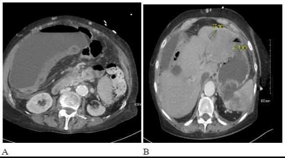
Case Description/Methods: A 72-year-old female, with a history of hypothyroidism, presented with non-specific upper abdominal pain, abdominal distension, fatigue, and 24 pounds weight loss over 6 months. Physical examination was notable for ascites without stigmata of chronic liver disease. Blood work revealed normal CBC, CMP, and tumor markers. A CT scan of the abdomen revealed multiple hypodense ill-defined masses throughout the liver with soft tissues fullness of the pancreas. A mass in the root of the mesentery, which appeared to be originating from the pancreas, was seen on MRI, along with focal fatty infiltration of the liver. Steatohepatitis was seen in the liver biopsy, while pancreatic mass biopsy revealed acute inflammation with areas of fibrosis but no indication of malignancy. Repeat EUS-FNB returned with nonmalignant findings. Ascitic fluid cytology was negative for malignancy. she has had multiple hospitalizations for Spontaneous bacterial peritonitis, intra abdominal venous thrombosis, ileus etc.
Repeated CT abdomen 4 months later, showed extensive desmoplastic reaction and fibrosis in the root of the mesentery, obliterating superior mesenteric vein and splenic vein producing cavernous transformation of the portal vein. ESR, CRP, and ANA were elevated. Serum IgG and IgG4 were elevated to 1723 and 156.9 respectively. ANA-specific antibodies, ANCA, and cardiolipin antibody were negative.IgG4 immunohistochemical analysis was performed however remained inconclusive with an average of 6 IgG4 and 22 IgG plasma cells per HPF due to insufficient sample tissue. The mass-forming lesion was diagnosed as IgG4-related retroperitoneal fibrosis. Treatment with prednisone was started with a very good clinical response. Follow-up CT a year later showed a stable mass. RPF-producing cavernous transformation of the portal vein, presenting as pancreatic mass, and portal hypertension have rarely been documented.
Discussion: RPF is a rare fibroinflammatory disorder affecting ~1.2 cases per 100,000 patients per year. CT, MRI, and PET are the mainstay of noninvasive diagnosis of the disease. Glucocorticoids are the mainstay of treatment.

Disclosures:
Rameela Mahat, MD1, Neelima Reddy, MD2. E0077 - A Diagnostic Conundrum: Pancreatic Retroperitoneal Fibrosis Masquerading as a Metastatic Pancreatic Tumor, ACG 2022 Annual Scientific Meeting Abstracts. Charlotte, NC: American College of Gastroenterology.
1Baton Rouge General Internal Medicine Residency Program, Baton Rouge, LA; 2Texas Digestive Disease Consultants, Baton Rouge, LA
Introduction: Retroperitoneal fibrosis (RPF) is a rare condition, characterized by inflammation and progressive development of fibrotic mass in the retroperitoneal space. Due to the wide variety of presentations, diagnosis remains challenging, difficult, and often delayed. This case highlights an unusual presentation of this rare entity
Case Description/Methods: A 72-year-old female, with a history of hypothyroidism, presented with non-specific upper abdominal pain, abdominal distension, fatigue, and 24 pounds weight loss over 6 months. Physical examination was notable for ascites without stigmata of chronic liver disease. Blood work revealed normal CBC, CMP, and tumor markers. A CT scan of the abdomen revealed multiple hypodense ill-defined masses throughout the liver with soft tissues fullness of the pancreas. A mass in the root of the mesentery, which appeared to be originating from the pancreas, was seen on MRI, along with focal fatty infiltration of the liver. Steatohepatitis was seen in the liver biopsy, while pancreatic mass biopsy revealed acute inflammation with areas of fibrosis but no indication of malignancy. Repeat EUS-FNB returned with nonmalignant findings. Ascitic fluid cytology was negative for malignancy. she has had multiple hospitalizations for Spontaneous bacterial peritonitis, intra abdominal venous thrombosis, ileus etc.
Repeated CT abdomen 4 months later, showed extensive desmoplastic reaction and fibrosis in the root of the mesentery, obliterating superior mesenteric vein and splenic vein producing cavernous transformation of the portal vein. ESR, CRP, and ANA were elevated. Serum IgG and IgG4 were elevated to 1723 and 156.9 respectively. ANA-specific antibodies, ANCA, and cardiolipin antibody were negative.IgG4 immunohistochemical analysis was performed however remained inconclusive with an average of 6 IgG4 and 22 IgG plasma cells per HPF due to insufficient sample tissue. The mass-forming lesion was diagnosed as IgG4-related retroperitoneal fibrosis. Treatment with prednisone was started with a very good clinical response. Follow-up CT a year later showed a stable mass. RPF-producing cavernous transformation of the portal vein, presenting as pancreatic mass, and portal hypertension have rarely been documented.
Discussion: RPF is a rare fibroinflammatory disorder affecting ~1.2 cases per 100,000 patients per year. CT, MRI, and PET are the mainstay of noninvasive diagnosis of the disease. Glucocorticoids are the mainstay of treatment.

Figure: Fig A: Infiltrative soft tissue density mass above the pancreas concerning malignancy with occlusion of the portal vein and SMV. Fig B: Indeterminate ill-defined hypodense hepatic lesions.
Disclosures:
Rameela Mahat indicated no relevant financial relationships.
Neelima Reddy indicated no relevant financial relationships.
Rameela Mahat, MD1, Neelima Reddy, MD2. E0077 - A Diagnostic Conundrum: Pancreatic Retroperitoneal Fibrosis Masquerading as a Metastatic Pancreatic Tumor, ACG 2022 Annual Scientific Meeting Abstracts. Charlotte, NC: American College of Gastroenterology.
