Back
Poster Session E - Tuesday Afternoon
E0161 - Streptococcus Bovis Bacteremia and Endocarditis Leading to the Diagnosis of Synchronous Colon and Appendiceal Adenocarcinoma
Tuesday, October 25, 2022
3:00 PM – 5:00 PM ET
Location: Crown Ballroom

Alicia Kovach, MD
Saint Michael's Medical Center
Newark, NJ
Presenting Author(s)
Alicia Kovach, MD1, Dema Shamoon, MD2, Andre Fedida, MD3, Jennifer Brown, DO3
1Saint Michael's Medical Center, Newark, NJ; 2Saint Michael's Medical Center, New York Medical College, Newark, NJ; 3St. Michael's Medical Center, New York Medical College, Newark, NJ
Introduction: Colon adenocarcinoma has been shown to be associated with appendiceal adenocarcinoma and Streptococcus bovis (S. bovis) infection. An association between appendiceal cancer and S. bovis infection has not been reported. This is a case of S. bovis bacteremia prompting colonoscopy and subsequent diagnosis of colon adenocarcinoma and incidental diagnosis of appendiceal adenocarcinoma.
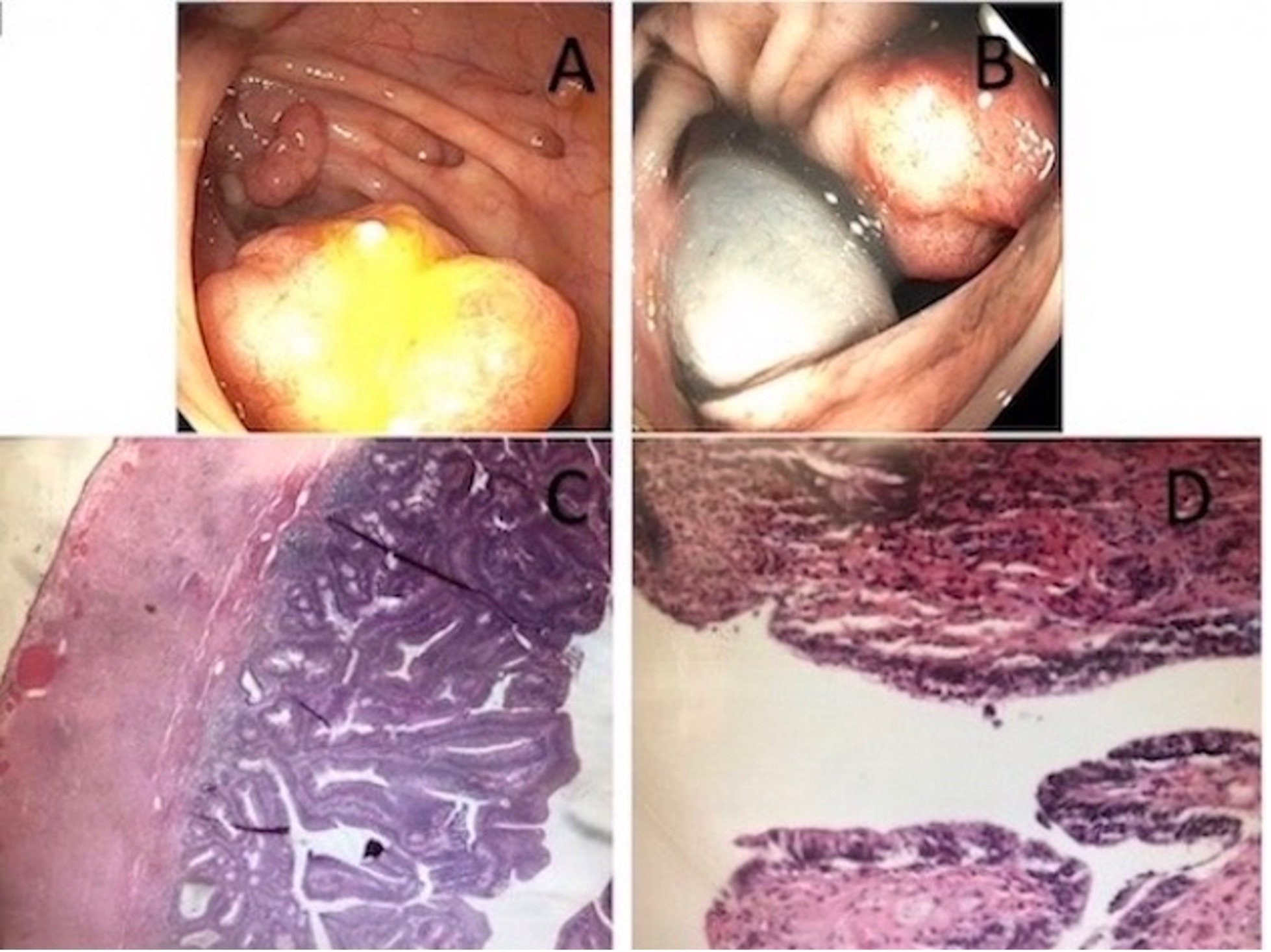
Case Description/Methods: A 77-year-old male with a history of metabolic syndrome, coronary artery bypass graft surgery, abdominal aortic aneurysm, heart failure with preserved ejection fraction, and aortic stenosis came to the emergency department for dyspnea and chest pressure. Vital signs were stable with a temperature of 103.2 °F. Physical exam revealed diffuse lung rales and bilateral lower extremity trace edema. Labs are attached (table 1). ECG was equivocal for acute myocardial infarction and cardiac catheterization was negative. Blood cultures grew S. bovis. TTE showed no valvular vegetations. TEE showed a mitral valve vegetation consistent with infective endocarditis (IE). Lower extremity vein thrombosis was diagnosed; anticoagulation was started which led to anemia and melena. Colonoscopy showed a large mass in the ascending colon (AC), a large villous appearing mass in the sigmoid colon (SC), multiple polyps, and severe diffuse diverticulosis (Images A, B). CT abdomen/pelvis showed no evidence of metastasis. He underwent SC and AC resections. Pathology of the SC showed tubulovillous adenoma with high-grade dysplasia and negative for submucosal invasion (Image C). Pathology of the AC showed well-differentiated appendiceal adenocarcinoma of the distal appendix invading the muscularis propria with appendiceal serosa and fat negative for invasion. Pathology of the AC also showed intramucosal adenocarcinoma without lymphovascular space invasion (Image D). Lymph nodes were negative for metastasis.
Discussion: Appendiceal cancer is rare. It affected only 2900 individuals in the US from 2012-2016. It is usually diagnosed after appendectomy for appendicitis. It has been proposed that patients with appendiceal adenocarcinoma carry an increased risk of colon adenocarcinoma and should be screened with colonoscopy at time of diagnosis. In this case, S. bovis bacteremia and IE prompted a colonoscopy. It remains unknown if there is an association between appendiceal adenocarcinoma and S. bovis infection. This report illustrates one case of concomitant colon and appendiceal adenocarcinoma with S. bovis bacteremia and IE.

Disclosures:
Alicia Kovach, MD1, Dema Shamoon, MD2, Andre Fedida, MD3, Jennifer Brown, DO3. E0161 - Streptococcus Bovis Bacteremia and Endocarditis Leading to the Diagnosis of Synchronous Colon and Appendiceal Adenocarcinoma, ACG 2022 Annual Scientific Meeting Abstracts. Charlotte, NC: American College of Gastroenterology.
1Saint Michael's Medical Center, Newark, NJ; 2Saint Michael's Medical Center, New York Medical College, Newark, NJ; 3St. Michael's Medical Center, New York Medical College, Newark, NJ
Introduction: Colon adenocarcinoma has been shown to be associated with appendiceal adenocarcinoma and Streptococcus bovis (S. bovis) infection. An association between appendiceal cancer and S. bovis infection has not been reported. This is a case of S. bovis bacteremia prompting colonoscopy and subsequent diagnosis of colon adenocarcinoma and incidental diagnosis of appendiceal adenocarcinoma.
Case Description/Methods: A 77-year-old male with a history of metabolic syndrome, coronary artery bypass graft surgery, abdominal aortic aneurysm, heart failure with preserved ejection fraction, and aortic stenosis came to the emergency department for dyspnea and chest pressure. Vital signs were stable with a temperature of 103.2 °F. Physical exam revealed diffuse lung rales and bilateral lower extremity trace edema. Labs are attached (table 1). ECG was equivocal for acute myocardial infarction and cardiac catheterization was negative. Blood cultures grew S. bovis. TTE showed no valvular vegetations. TEE showed a mitral valve vegetation consistent with infective endocarditis (IE). Lower extremity vein thrombosis was diagnosed; anticoagulation was started which led to anemia and melena. Colonoscopy showed a large mass in the ascending colon (AC), a large villous appearing mass in the sigmoid colon (SC), multiple polyps, and severe diffuse diverticulosis (Images A, B). CT abdomen/pelvis showed no evidence of metastasis. He underwent SC and AC resections. Pathology of the SC showed tubulovillous adenoma with high-grade dysplasia and negative for submucosal invasion (Image C). Pathology of the AC showed well-differentiated appendiceal adenocarcinoma of the distal appendix invading the muscularis propria with appendiceal serosa and fat negative for invasion. Pathology of the AC also showed intramucosal adenocarcinoma without lymphovascular space invasion (Image D). Lymph nodes were negative for metastasis.
Discussion: Appendiceal cancer is rare. It affected only 2900 individuals in the US from 2012-2016. It is usually diagnosed after appendectomy for appendicitis. It has been proposed that patients with appendiceal adenocarcinoma carry an increased risk of colon adenocarcinoma and should be screened with colonoscopy at time of diagnosis. In this case, S. bovis bacteremia and IE prompted a colonoscopy. It remains unknown if there is an association between appendiceal adenocarcinoma and S. bovis infection. This report illustrates one case of concomitant colon and appendiceal adenocarcinoma with S. bovis bacteremia and IE.

Figure: Image A: Colonoscopy image showing ascending colon mass with diverticulosis and other polyps.
Image B: Colonoscopy image showing sigmoid colon mass.
Image C: Histological image of sigmoid colon resection showing tubulovillous adenoma with high-grade dysplasia.
Image D: Histological image of right hemicolectomy specimen showing in-situ adenocarcinoma.
Image B: Colonoscopy image showing sigmoid colon mass.
Image C: Histological image of sigmoid colon resection showing tubulovillous adenoma with high-grade dysplasia.
Image D: Histological image of right hemicolectomy specimen showing in-situ adenocarcinoma.
| Laboratory test | Value | Reference range |
| Troponin I | 4.270 | 0.000-0.450 ng/mL |
| Lactic acid | 2.2 | 0-2 mmol/L |
| WBC | 9.00 | 4.40-11.00 103/μL |
Table: Table 1: Laboratory Values.
Disclosures:
Alicia Kovach indicated no relevant financial relationships.
Dema Shamoon indicated no relevant financial relationships.
Andre Fedida indicated no relevant financial relationships.
Jennifer Brown indicated no relevant financial relationships.
Alicia Kovach, MD1, Dema Shamoon, MD2, Andre Fedida, MD3, Jennifer Brown, DO3. E0161 - Streptococcus Bovis Bacteremia and Endocarditis Leading to the Diagnosis of Synchronous Colon and Appendiceal Adenocarcinoma, ACG 2022 Annual Scientific Meeting Abstracts. Charlotte, NC: American College of Gastroenterology.
