Back


Poster Session A - Sunday Afternoon
Category: GI Bleeding
A0323 - Successfully Treated Severe Cameron Type Ulcer in a Recurrent Morgagni Hernia: A Case Report
Sunday, October 23, 2022
5:00 PM – 7:00 PM ET
Location: Crown Ballroom

Has Audio

Brian Sowka, DO
Gundersen Health System
La Crosse, WI
Presenting Author(s)
Brian Sowka, DO, Padmavathi Mali, MD, Milan Folkers, MD
Gundersen Health System, La Crosse, WI
Introduction: Morgagni hernias are rare diaphragmatic hernias located in the anterior or retrosternal areas and comprise only 2-5% of congenital hernias. There are only a handful of cases in the literature in which these hernias present with upper gastrointestinal bleeding. Typical endoscopic treatments can be limited due to anatomic and technical reasons. We present a patient with successfully treated upper gastrointestinal bleed from a recurrent Morgagni hernia.
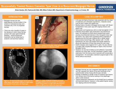
Case Description/Methods: A 44-year-old Native American male presented with four days of weakness and orthostatic hypotension to the emergency department. Medical history included a Morgagni hernia repair with Nissen fundoplication many years ago with ongoing tobacco and aspirin use. He had three melanotic stools over the preceding two weeks along with a 50-pound unintentional weight loss over the last year. He was tachycardic and hypotensive with an initial hemoglobin of 5.3. Following resuscitation with transfusions and intravenous fluids, he underwent an EGD which showed three ulcers causing circumferential stenosis in the mid-gastric body near the hernia. Two ulcers were superficial without stigmata of bleeding and the third was cratered with a visible vessel without active hemorrhage. After injection with 1:10,000 epinephrine, the ulcer was treated with bipolar diathermy along with placement of three clips along the ulcer base. Gastric biopsies were negative for H. pylori. He was referred to surgery after hospital discharge for repair of the recurrent Morgagni hernia. He was lost to follow-up and suffered a myocardial infarction two years later. He developed a gastric outlet obstruction from the hernia which improved with conservative management. He eventually underwent surgical repair of the hernia without recurrence.
Discussion: Morgagni hernias are rare congenital diaphragmatic hernias. These hernias usually develop on the right side of the diaphragm and can be found incidentally during chest or upper abdominal imaging. They originate from failure of the pars tendinalis of the costochondral arches fusing with the pars sternalis. The underlying pathophysiology of bleeding is similar to that of Cameron type lesions from erosion due to the hernia rubbing the diaphragm defect. Typical endoscopic treatments can be limited due to anatomic and technical reasons, however, in this patient bipolar cautery and clipping achieved hemostasis. Definitive management of Morgagni hernias include surgery to prevent complications including incarceration.

Disclosures:
Brian Sowka, DO, Padmavathi Mali, MD, Milan Folkers, MD. A0323 - Successfully Treated Severe Cameron Type Ulcer in a Recurrent Morgagni Hernia: A Case Report, ACG 2022 Annual Scientific Meeting Abstracts. Charlotte, NC: American College of Gastroenterology.
Gundersen Health System, La Crosse, WI
Introduction: Morgagni hernias are rare diaphragmatic hernias located in the anterior or retrosternal areas and comprise only 2-5% of congenital hernias. There are only a handful of cases in the literature in which these hernias present with upper gastrointestinal bleeding. Typical endoscopic treatments can be limited due to anatomic and technical reasons. We present a patient with successfully treated upper gastrointestinal bleed from a recurrent Morgagni hernia.
Case Description/Methods: A 44-year-old Native American male presented with four days of weakness and orthostatic hypotension to the emergency department. Medical history included a Morgagni hernia repair with Nissen fundoplication many years ago with ongoing tobacco and aspirin use. He had three melanotic stools over the preceding two weeks along with a 50-pound unintentional weight loss over the last year. He was tachycardic and hypotensive with an initial hemoglobin of 5.3. Following resuscitation with transfusions and intravenous fluids, he underwent an EGD which showed three ulcers causing circumferential stenosis in the mid-gastric body near the hernia. Two ulcers were superficial without stigmata of bleeding and the third was cratered with a visible vessel without active hemorrhage. After injection with 1:10,000 epinephrine, the ulcer was treated with bipolar diathermy along with placement of three clips along the ulcer base. Gastric biopsies were negative for H. pylori. He was referred to surgery after hospital discharge for repair of the recurrent Morgagni hernia. He was lost to follow-up and suffered a myocardial infarction two years later. He developed a gastric outlet obstruction from the hernia which improved with conservative management. He eventually underwent surgical repair of the hernia without recurrence.
Discussion: Morgagni hernias are rare congenital diaphragmatic hernias. These hernias usually develop on the right side of the diaphragm and can be found incidentally during chest or upper abdominal imaging. They originate from failure of the pars tendinalis of the costochondral arches fusing with the pars sternalis. The underlying pathophysiology of bleeding is similar to that of Cameron type lesions from erosion due to the hernia rubbing the diaphragm defect. Typical endoscopic treatments can be limited due to anatomic and technical reasons, however, in this patient bipolar cautery and clipping achieved hemostasis. Definitive management of Morgagni hernias include surgery to prevent complications including incarceration.

Figure: A - Vessel oozing after initial contact in stomach view prior to clipping on EGD
B - Upper GI Series showing recurrent Morgagni Hernia with first portion of duodenum above hernia defect on right side with stomach body and GE junction below the diaphragm
C - CT showing Morgagni hernia prior to initial repair with stomach antrum in right chest and stomach body under left diaphragm
Disclosures:
Brian Sowka indicated no relevant financial relationships.
Padmavathi Mali indicated no relevant financial relationships.
Milan Folkers indicated no relevant financial relationships.
Brian Sowka, DO, Padmavathi Mali, MD, Milan Folkers, MD. A0323 - Successfully Treated Severe Cameron Type Ulcer in a Recurrent Morgagni Hernia: A Case Report, ACG 2022 Annual Scientific Meeting Abstracts. Charlotte, NC: American College of Gastroenterology.
