Back


Poster Session A - Sunday Afternoon
Category: Interventional Endoscopy
A0432 - Endoscopic Management of an Obstructing Duodenal Wall Hematoma in the Setting of Acute Pancreatitis
Sunday, October 23, 2022
5:00 PM – 7:00 PM ET
Location: Crown Ballroom

Has Audio

Jordan K. Voss, MD
Mayo Clinic
Rochester, MN
Presenting Author(s)
Jordan K. Voss, MD, Jissy Cyriac, MD, Aliana M. Bofill-Garcia, MD, Raseen Tariq, MD, Vinay Chandrasekhara, MD, Ryan Law, DO, Conor Loftus, MD
Mayo Clinic, Rochester, MN
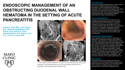
Introduction: Spontaneous duodenal wall hematoma is a rare condition which can be associated with acute and chronic pancreatitis. It is often complicated by small bowel obstruction. When conservative management fails, options are limited. Here we describe a case of small bowel obstruction secondary to pancreatitis-associated duodenal wall hematoma which was successfully managed endoscopically through cystduodenostomy.
Case Description/Methods: A 69-year-old male with HIV and normal CD4 count presented with acute abdominal pain and non-bloody emesis with elevated lipase of 374 U/L (ULN 82).
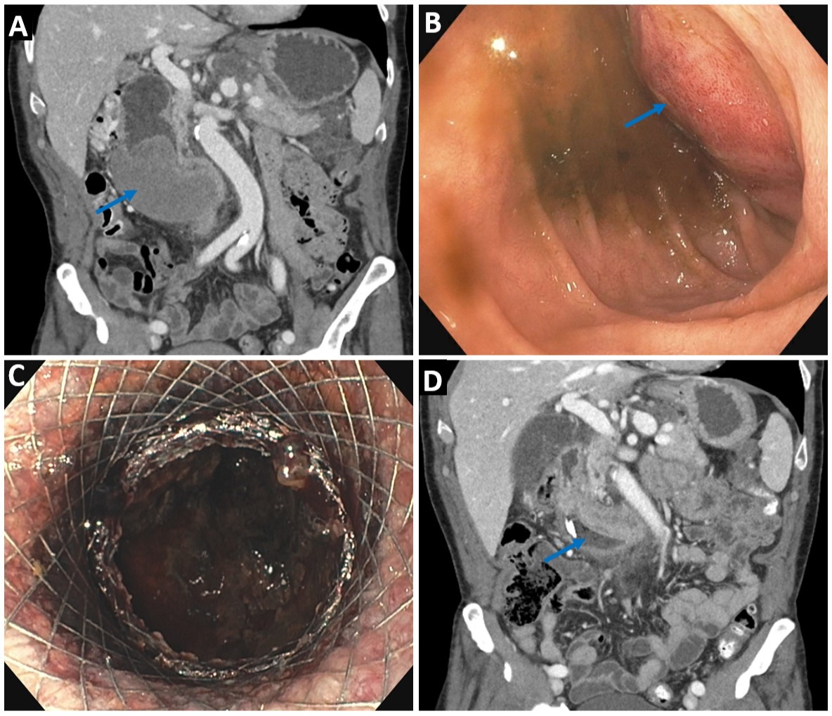
Computed tomography revealed a 5.8 cm multilobulated pancreatic pseudocyst with associated inflammation and coarse calcifications and a 10 cm circumferential submucosal mass within the duodenum with significant luminal narrowing and compression of the adjacent inferior vena cava (a). Endoscopic ultrasound confirmed a homogenous, hypoechoic subepithelial lesion obstructing the second portion of the duodenum consistent with a hematoma (b). A nasogastric-jejunal tube was placed for feeding and decompression.
Due to persistent obstruction and IVC compression, on hospital day 10 endoscopic drainage of the duodenal hematoma was performed via cystduodenostomy with a 15x10 mm electrocautery-enhanced lumen-apposing metal stent (LAMS) (AXIOS, Boston Scientific, Marlborough, MA, USA) (c). The LAMS was immediately removed after near-complete evacuation of the hematoma cavity to minimize the risk of bleeding complications. A plastic double-pigtail stent was left in place to facilitate complete drainage. The duodenum was then dilated with a 12-13.5-15mm hydrostatic balloon dilator. The pigtail stent was removed 3 days later with clinical, radiographic, and endoscopic resolution of the duodenal obstruction (d).
Discussion: Duodenal hematoma is a previously described rare complication of pancreatitis. Endoscopic evacuation was a safe and effective means of relieving the duodenal obstruction in this patient. This minimally invasive option resulted in immediate resolution and can be considered when conservative management fails.

Disclosures:
Jordan K. Voss, MD, Jissy Cyriac, MD, Aliana M. Bofill-Garcia, MD, Raseen Tariq, MD, Vinay Chandrasekhara, MD, Ryan Law, DO, Conor Loftus, MD. A0432 - Endoscopic Management of an Obstructing Duodenal Wall Hematoma in the Setting of Acute Pancreatitis, ACG 2022 Annual Scientific Meeting Abstracts. Charlotte, NC: American College of Gastroenterology.
Mayo Clinic, Rochester, MN
Introduction: Spontaneous duodenal wall hematoma is a rare condition which can be associated with acute and chronic pancreatitis. It is often complicated by small bowel obstruction. When conservative management fails, options are limited. Here we describe a case of small bowel obstruction secondary to pancreatitis-associated duodenal wall hematoma which was successfully managed endoscopically through cystduodenostomy.
Case Description/Methods: A 69-year-old male with HIV and normal CD4 count presented with acute abdominal pain and non-bloody emesis with elevated lipase of 374 U/L (ULN 82).
Computed tomography revealed a 5.8 cm multilobulated pancreatic pseudocyst with associated inflammation and coarse calcifications and a 10 cm circumferential submucosal mass within the duodenum with significant luminal narrowing and compression of the adjacent inferior vena cava (a). Endoscopic ultrasound confirmed a homogenous, hypoechoic subepithelial lesion obstructing the second portion of the duodenum consistent with a hematoma (b). A nasogastric-jejunal tube was placed for feeding and decompression.
Due to persistent obstruction and IVC compression, on hospital day 10 endoscopic drainage of the duodenal hematoma was performed via cystduodenostomy with a 15x10 mm electrocautery-enhanced lumen-apposing metal stent (LAMS) (AXIOS, Boston Scientific, Marlborough, MA, USA) (c). The LAMS was immediately removed after near-complete evacuation of the hematoma cavity to minimize the risk of bleeding complications. A plastic double-pigtail stent was left in place to facilitate complete drainage. The duodenum was then dilated with a 12-13.5-15mm hydrostatic balloon dilator. The pigtail stent was removed 3 days later with clinical, radiographic, and endoscopic resolution of the duodenal obstruction (d).
Discussion: Duodenal hematoma is a previously described rare complication of pancreatitis. Endoscopic evacuation was a safe and effective means of relieving the duodenal obstruction in this patient. This minimally invasive option resulted in immediate resolution and can be considered when conservative management fails.

Figure: 10 cm duodenal wall hematoma on CT scan (a) and endoscopy (b). Endoscopic drainage via cystduodenostomy with a lumen-apposing metal stent (c). Resolution of the duodenal wall hematoma (d).
Disclosures:
Jordan Voss indicated no relevant financial relationships.
Jissy Cyriac indicated no relevant financial relationships.
Aliana Bofill-Garcia indicated no relevant financial relationships.
Raseen Tariq indicated no relevant financial relationships.
Vinay Chandrasekhara: Boston Scientific – Consultant. Covidien, LP – Consultant. Nevakar Corporation – Stock-privately held company.
Ryan Law: Boston Scientific – Consultant. ConMed – Consultant. Medtronic – Consultant. Olympus – Consultant. UpToDate – Royalties.
Conor Loftus indicated no relevant financial relationships.
Jordan K. Voss, MD, Jissy Cyriac, MD, Aliana M. Bofill-Garcia, MD, Raseen Tariq, MD, Vinay Chandrasekhara, MD, Ryan Law, DO, Conor Loftus, MD. A0432 - Endoscopic Management of an Obstructing Duodenal Wall Hematoma in the Setting of Acute Pancreatitis, ACG 2022 Annual Scientific Meeting Abstracts. Charlotte, NC: American College of Gastroenterology.

