Back


Poster Session B - Monday Morning
Category: Biliary/Pancreas
B0043 - Neuroendocrine Tumor in the Biliary Tract Presenting With Cholangitis
Monday, October 24, 2022
10:00 AM – 12:00 PM ET
Location: Crown Ballroom

Has Audio

Eugene C. Nwankwo, Jr., MD
Saint Louis University
St. Louis, MO
Presenting Author(s)
Eugene C. Nwankwo, MD1, Gebran Khneizer, MD1, Gregory Sayuk, MD2, Michael Presti, MD3, Jill E. Elwing, MD2
1Saint Louis University, St. Louis, MO; 2St. Louis VA, Washington University, St. Louis, MO; 3St. Louis VA, Saint Louis University, St. Louis, MO
Introduction: Neuroendocrine tumors (NETs) of the biliary tract account for less than 2% of all gastrointestinal cancers with an incidence of 0.32%.1,2 Extrahepatic biliary NETs are mostly found incidentally in the distal common bile duct (CBD) but in rare cases they may present with obstructive jaundice. We present a case of an elderly male with symptoms of biliary obstruction and acute cholangitis secondary to a NET in the proximal bile duct.
Case Description/Methods:
A 64-year-old male with hypertension presented with epigastric abdominal pain, vomiting and fever. Patient reported an unintentional 50-pound weight loss over the past year. Vital signs were remarkable for heart rate of 118 beats/min. Labs revealed white blood cell count of 21,000 uL, AST 303 U/L, ALT 563 U/L, alkaline phosphatase 300 U/L, total bilirubin 1.9 mg/dL.
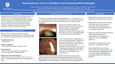
CT abdomen showed intrahepatic ductal dilatation to 1.1 cm, and a 1.2 x 1.1 cm low attenuating lesion within the left lobe of the liver and wall thickening of the proximal jejunal loops. Piperacillin-tazobactam was started for acute cholangitis. Endoscopic ultrasound showed duct dilation in the left hepatic lobe and ERCP showed ulceration of major papillae. Following biliary sphincterotomy, exploration revealed severe stenosis of the main bile duct and a 12 mm polypoid lesion in the upper third of the CBD. Histology showed well differentiated NET with trabecular and organoid pattern, positive chromogranin and synaptophysin with Ki-67 index of 3%. Special staining was also positive for reticulin and trichrome.
A 10fr x 9cm stent was placed. Following a 7-day course of antibiotics and symptom resolution, the patient was discharged with a plan for elective surgery. Left hepatectomy, CBD resection, and Roux-en-Y hepaticojejunostomy were performed 6 months after hospitalization. At 2 months follow up in clinic, the patient was asymptomatic.
Discussion: Biliary NETs are found in the common hepatic duct, CBD and the cystic duct.3 Biliary NETs are twice more common in females, with a mean age of 47 years old. Although most cases are incidental, presenting symptoms are jaundice and pruritis. Diagnosis requires histologic evidence and positive expression of chromogranin A, synaptophysin, CD56, or neural cell adhesion molecule.4 Treatment is surgical excision with Roux-en-Y hepaticojejunostomy. Since biliary NETs uncommonly present with obstructive jaundice, this case presentation highlights the need for a broad differential diagnosis in evaluating patients with obstructive jaundice.
Disclosures:
Eugene C. Nwankwo, MD1, Gebran Khneizer, MD1, Gregory Sayuk, MD2, Michael Presti, MD3, Jill E. Elwing, MD2. B0043 - Neuroendocrine Tumor in the Biliary Tract Presenting With Cholangitis, ACG 2022 Annual Scientific Meeting Abstracts. Charlotte, NC: American College of Gastroenterology.
1Saint Louis University, St. Louis, MO; 2St. Louis VA, Washington University, St. Louis, MO; 3St. Louis VA, Saint Louis University, St. Louis, MO
Introduction: Neuroendocrine tumors (NETs) of the biliary tract account for less than 2% of all gastrointestinal cancers with an incidence of 0.32%.1,2 Extrahepatic biliary NETs are mostly found incidentally in the distal common bile duct (CBD) but in rare cases they may present with obstructive jaundice. We present a case of an elderly male with symptoms of biliary obstruction and acute cholangitis secondary to a NET in the proximal bile duct.
Case Description/Methods:
A 64-year-old male with hypertension presented with epigastric abdominal pain, vomiting and fever. Patient reported an unintentional 50-pound weight loss over the past year. Vital signs were remarkable for heart rate of 118 beats/min. Labs revealed white blood cell count of 21,000 uL, AST 303 U/L, ALT 563 U/L, alkaline phosphatase 300 U/L, total bilirubin 1.9 mg/dL.
CT abdomen showed intrahepatic ductal dilatation to 1.1 cm, and a 1.2 x 1.1 cm low attenuating lesion within the left lobe of the liver and wall thickening of the proximal jejunal loops. Piperacillin-tazobactam was started for acute cholangitis. Endoscopic ultrasound showed duct dilation in the left hepatic lobe and ERCP showed ulceration of major papillae. Following biliary sphincterotomy, exploration revealed severe stenosis of the main bile duct and a 12 mm polypoid lesion in the upper third of the CBD. Histology showed well differentiated NET with trabecular and organoid pattern, positive chromogranin and synaptophysin with Ki-67 index of 3%. Special staining was also positive for reticulin and trichrome.
A 10fr x 9cm stent was placed. Following a 7-day course of antibiotics and symptom resolution, the patient was discharged with a plan for elective surgery. Left hepatectomy, CBD resection, and Roux-en-Y hepaticojejunostomy were performed 6 months after hospitalization. At 2 months follow up in clinic, the patient was asymptomatic.
Discussion: Biliary NETs are found in the common hepatic duct, CBD and the cystic duct.3 Biliary NETs are twice more common in females, with a mean age of 47 years old. Although most cases are incidental, presenting symptoms are jaundice and pruritis. Diagnosis requires histologic evidence and positive expression of chromogranin A, synaptophysin, CD56, or neural cell adhesion molecule.4 Treatment is surgical excision with Roux-en-Y hepaticojejunostomy. Since biliary NETs uncommonly present with obstructive jaundice, this case presentation highlights the need for a broad differential diagnosis in evaluating patients with obstructive jaundice.
Disclosures:
Eugene Nwankwo indicated no relevant financial relationships.
Gebran Khneizer indicated no relevant financial relationships.
Gregory Sayuk indicated no relevant financial relationships.
Michael Presti indicated no relevant financial relationships.
Jill Elwing indicated no relevant financial relationships.
Eugene C. Nwankwo, MD1, Gebran Khneizer, MD1, Gregory Sayuk, MD2, Michael Presti, MD3, Jill E. Elwing, MD2. B0043 - Neuroendocrine Tumor in the Biliary Tract Presenting With Cholangitis, ACG 2022 Annual Scientific Meeting Abstracts. Charlotte, NC: American College of Gastroenterology.
