Back


Poster Session B - Monday Morning
Category: Biliary/Pancreas
B0072 - Obstructive Jaundice From Adenosquamous Carcinoma of the Ampulla of Vater
Monday, October 24, 2022
10:00 AM – 12:00 PM ET
Location: Crown Ballroom

Has Audio
.jpg)
Mark Hsu, MD
Kirk Kerkorian School of Medicine at UNLV
Las Vegas, NV
Presenting Author(s)
Mark Hsu, MD, Katrina Naik, MD, Amith Subhash, MD, Jose Aponte-Pieras, MD, Annie Hong, MD, Yousif Elmofti, MD, Muhammad Talha Farooqi, MD, Shahid Wahid, MD
Kirk Kerkorian School of Medicine at UNLV, Las Vegas, NV
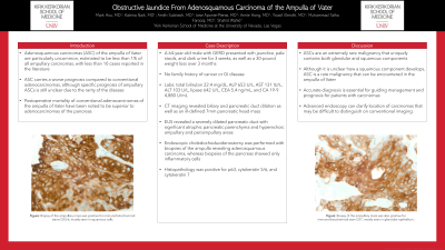
Introduction: Adenosquamous carcinoma of the pancreatobiliary system is a rare malignancy of the gastrointestinal tract, and uniquely unusual due to its composition of both glandular and squamous components. While it carries a worse prognosis compared to more common adenocarcinomas, adenosquamous carcinoma of the ampulla of Vater is reported to have superior postoperative mortality compared to that of the pancreas. Thus, accurate diagnosis is essential for guiding management and prognosis for the patient. Here we report a case of ampullary adenosquamous carcinoma presenting as obstructive jaundice, with imaging suggestive of a pancreatic mass but later identified as ampullary by advanced endoscopy.
Case Description/Methods: A 64-year-old male with GERD presented with 3 weeks of jaundice, pale stools, and dark urine. He was noted to have a 30-pound weight loss over 3 months. He denied a family history of cancer or gastrointestinal disease. The following labs were noted: total bilirubin 22.4 mg/dL, ALP 653 U/L, AST 131 IU/L, ALT 103 U/L, lipase 642 U/L, CEA 5.4 ng/mL, and CA 19-9 4,880 U/mL. CT imaging revealed biliary and pancreatic duct dilation, as well as an ill-defined 7mm pancreatic head mass. EUS revealed a severely dilated pancreatic duct with significant atrophic pancreatic parenchyma and hyperechoic ampullary and periampullary areas. A choledochoduodenostomy was performed with biopsies of the ampulla revealing adenosquamous carcinoma, whereas biopsies of the pancreas showed only inflammatory cells. Histopathology was positive for cytokeratin 5 and 6, focally positive for cytokeratin 7, and positive for p63, which are similar to stains utilized in adenosquamous carcinoma of the pancreas.
Discussion: While conventional imaging initially suggested a pancreatic malignancy for our patient, advanced endoscopy revealed a rare ampullary malignancy. The histopathology of this patient’s adenosquamous carcinoma of the ampulla of Vater resembled that of adenosquamous carcinoma of the pancreas. Postoperative mortality for the former, however, can be more favorable than the latter. This case highlights the importance of accurate distinction of pancreatobiliary adenosquamous carcinoma, and underscores advanced endoscopy as a valuable tool for achieving the correct diagnosis.

Disclosures:
Mark Hsu, MD, Katrina Naik, MD, Amith Subhash, MD, Jose Aponte-Pieras, MD, Annie Hong, MD, Yousif Elmofti, MD, Muhammad Talha Farooqi, MD, Shahid Wahid, MD. B0072 - Obstructive Jaundice From Adenosquamous Carcinoma of the Ampulla of Vater, ACG 2022 Annual Scientific Meeting Abstracts. Charlotte, NC: American College of Gastroenterology.
Kirk Kerkorian School of Medicine at UNLV, Las Vegas, NV
Introduction: Adenosquamous carcinoma of the pancreatobiliary system is a rare malignancy of the gastrointestinal tract, and uniquely unusual due to its composition of both glandular and squamous components. While it carries a worse prognosis compared to more common adenocarcinomas, adenosquamous carcinoma of the ampulla of Vater is reported to have superior postoperative mortality compared to that of the pancreas. Thus, accurate diagnosis is essential for guiding management and prognosis for the patient. Here we report a case of ampullary adenosquamous carcinoma presenting as obstructive jaundice, with imaging suggestive of a pancreatic mass but later identified as ampullary by advanced endoscopy.
Case Description/Methods: A 64-year-old male with GERD presented with 3 weeks of jaundice, pale stools, and dark urine. He was noted to have a 30-pound weight loss over 3 months. He denied a family history of cancer or gastrointestinal disease. The following labs were noted: total bilirubin 22.4 mg/dL, ALP 653 U/L, AST 131 IU/L, ALT 103 U/L, lipase 642 U/L, CEA 5.4 ng/mL, and CA 19-9 4,880 U/mL. CT imaging revealed biliary and pancreatic duct dilation, as well as an ill-defined 7mm pancreatic head mass. EUS revealed a severely dilated pancreatic duct with significant atrophic pancreatic parenchyma and hyperechoic ampullary and periampullary areas. A choledochoduodenostomy was performed with biopsies of the ampulla revealing adenosquamous carcinoma, whereas biopsies of the pancreas showed only inflammatory cells. Histopathology was positive for cytokeratin 5 and 6, focally positive for cytokeratin 7, and positive for p63, which are similar to stains utilized in adenosquamous carcinoma of the pancreas.
Discussion: While conventional imaging initially suggested a pancreatic malignancy for our patient, advanced endoscopy revealed a rare ampullary malignancy. The histopathology of this patient’s adenosquamous carcinoma of the ampulla of Vater resembled that of adenosquamous carcinoma of the pancreas. Postoperative mortality for the former, however, can be more favorable than the latter. This case highlights the importance of accurate distinction of pancreatobiliary adenosquamous carcinoma, and underscores advanced endoscopy as a valuable tool for achieving the correct diagnosis.

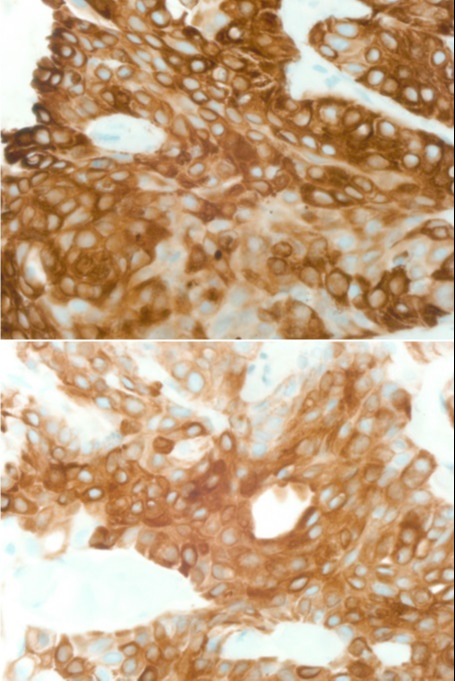
Figure: Figure: (Top) Biopsy of the ampullary mass was positive for immunohistochemical stains CK5/6, mostly seen in squamous cells. (Bottom) Biopsy of the ampullary mass was also positive for immunohistochemical stain CK7, mostly seen in glandular epithelium.
Disclosures:
Mark Hsu indicated no relevant financial relationships.
Katrina Naik indicated no relevant financial relationships.
Amith Subhash indicated no relevant financial relationships.
Jose Aponte-Pieras indicated no relevant financial relationships.
Annie Hong indicated no relevant financial relationships.
Yousif Elmofti indicated no relevant financial relationships.
Muhammad Talha Farooqi indicated no relevant financial relationships.
Shahid Wahid indicated no relevant financial relationships.
Mark Hsu, MD, Katrina Naik, MD, Amith Subhash, MD, Jose Aponte-Pieras, MD, Annie Hong, MD, Yousif Elmofti, MD, Muhammad Talha Farooqi, MD, Shahid Wahid, MD. B0072 - Obstructive Jaundice From Adenosquamous Carcinoma of the Ampulla of Vater, ACG 2022 Annual Scientific Meeting Abstracts. Charlotte, NC: American College of Gastroenterology.
