Back


Poster Session A - Sunday Afternoon
Category: Colon
A0125 - Extramammary Perianal Paget Disease, Colonoscopy Findings That Led to the Diagnosis of a Genitourinary Mass
Sunday, October 23, 2022
5:00 PM – 7:00 PM ET
Location: Crown Ballroom

Has Audio

Judie Hoilat, MD
Loyola University Medical Center
Maywood, IL
Presenting Author(s)
Award: Presidential Poster Award
Judie Hoilat, MD, Marcel Ghanim, MD, Sean McBrayer, MD, Zuie Wakade, DO, Abdul Haseeb, MD
Loyola University Medical Center, Maywood, IL
Introduction: Extramammary Paget’s disease (EMPD) is a rare cutaneous malignancy most commonly affecting the genitals, perineum, and perianal area. EMPD has cellular similarities to Paget’s breast cancer. It is difficult to estimate the true incidence of perianal Paget disease (PPD) due to its rarity but it is thought to represent 1.3% of all cases of Paget disease. We present a case of a 75-year-old Caucasian male who underwent a screening colonoscopy and was found to have perianal dermatitis which was biopsies and found to be PPD.
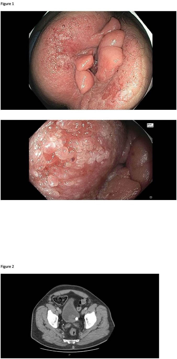
Case Description/Methods: A 75 year old Caucasian male underwent a colonoscopy for surveillance due to history of tubular adenomatous polyps. Eleven sub-centimeter polyps were resected, none of which showed evidence of colorectal adenocarcinoma. During the procedure, perianal exam revealed irregular circumferential erythematous rash. He was referred to colorectal surgery who performed a perianal skin biopsy which revealed extramammary Paget's disease. A computerized tomography of the chest, abdomen and pelvis was performed due to concern for secondary Paget's disease which revealed a calcified lesion in the left vesico-ureteral junction for which he will undergo evaluation by urology due to concern for malignancy.
Discussion: This case highlights the importance of careful examination of the perianal region during colonoscopy and the importance to maintain EMPD among the differential diagnosis for perianal dermatitis. There are two types of PPD; primary PPD represents carcinoma in situ of the apocrine gland ducts, whereas secondary PPD is thought to occur from intraepithelial spread of a separate underlying carcinoma.
Clinically patients may present with anal pain or pruritus, but some may be asymptomatic, as shown in our case. On examination, it appears as a slow-growing, erythematous plaque in the perianal region. PPD has been associated with synchronous or metachronous genitourinary and/or gastrointestinal malignancies. It is recommended that patients undergo screening for these malignancies but the appropriate frequency of surveillance remains unknown. When confirmed on biopsy, clinicians should closely monitor PPD for local progression and screen all patients for distant and local neoplasms.

Disclosures:
Judie Hoilat, MD, Marcel Ghanim, MD, Sean McBrayer, MD, Zuie Wakade, DO, Abdul Haseeb, MD. A0125 - Extramammary Perianal Paget Disease, Colonoscopy Findings That Led to the Diagnosis of a Genitourinary Mass, ACG 2022 Annual Scientific Meeting Abstracts. Charlotte, NC: American College of Gastroenterology.
Judie Hoilat, MD, Marcel Ghanim, MD, Sean McBrayer, MD, Zuie Wakade, DO, Abdul Haseeb, MD
Loyola University Medical Center, Maywood, IL
Introduction: Extramammary Paget’s disease (EMPD) is a rare cutaneous malignancy most commonly affecting the genitals, perineum, and perianal area. EMPD has cellular similarities to Paget’s breast cancer. It is difficult to estimate the true incidence of perianal Paget disease (PPD) due to its rarity but it is thought to represent 1.3% of all cases of Paget disease. We present a case of a 75-year-old Caucasian male who underwent a screening colonoscopy and was found to have perianal dermatitis which was biopsies and found to be PPD.
Case Description/Methods: A 75 year old Caucasian male underwent a colonoscopy for surveillance due to history of tubular adenomatous polyps. Eleven sub-centimeter polyps were resected, none of which showed evidence of colorectal adenocarcinoma. During the procedure, perianal exam revealed irregular circumferential erythematous rash. He was referred to colorectal surgery who performed a perianal skin biopsy which revealed extramammary Paget's disease. A computerized tomography of the chest, abdomen and pelvis was performed due to concern for secondary Paget's disease which revealed a calcified lesion in the left vesico-ureteral junction for which he will undergo evaluation by urology due to concern for malignancy.
Discussion: This case highlights the importance of careful examination of the perianal region during colonoscopy and the importance to maintain EMPD among the differential diagnosis for perianal dermatitis. There are two types of PPD; primary PPD represents carcinoma in situ of the apocrine gland ducts, whereas secondary PPD is thought to occur from intraepithelial spread of a separate underlying carcinoma.
Clinically patients may present with anal pain or pruritus, but some may be asymptomatic, as shown in our case. On examination, it appears as a slow-growing, erythematous plaque in the perianal region. PPD has been associated with synchronous or metachronous genitourinary and/or gastrointestinal malignancies. It is recommended that patients undergo screening for these malignancies but the appropriate frequency of surveillance remains unknown. When confirmed on biopsy, clinicians should closely monitor PPD for local progression and screen all patients for distant and local neoplasms.

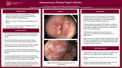
Figure: Figure 1 Perianal erythematous rash and hemorrhoids
Figure 2 Left calcified mass at vesico-ureteral junction measuring 1.3 cm
Figure 2 Left calcified mass at vesico-ureteral junction measuring 1.3 cm
Disclosures:
Judie Hoilat indicated no relevant financial relationships.
Marcel Ghanim indicated no relevant financial relationships.
Sean McBrayer indicated no relevant financial relationships.
Zuie Wakade indicated no relevant financial relationships.
Abdul Haseeb indicated no relevant financial relationships.
Judie Hoilat, MD, Marcel Ghanim, MD, Sean McBrayer, MD, Zuie Wakade, DO, Abdul Haseeb, MD. A0125 - Extramammary Perianal Paget Disease, Colonoscopy Findings That Led to the Diagnosis of a Genitourinary Mass, ACG 2022 Annual Scientific Meeting Abstracts. Charlotte, NC: American College of Gastroenterology.


