Back


Poster Session B - Monday Morning
Category: Biliary/Pancreas
B0034 - Choledochal Cyst Mimicking the Gallbladder With a Surprising Twist
Monday, October 24, 2022
10:00 AM – 12:00 PM ET
Location: Crown Ballroom

Has Audio

Sonia Samuel, DO
Albany Medical Center
Albany, NY
Presenting Author(s)
Sonia Samuel, DO, Umer Ejaz Malik, MD, Domenico Viterbo, MD
Albany Medical Center, Albany, NY
Introduction: Choledochal cysts are congenital rarities and often a pediatric diagnosis but 20% go undetected until adulthood. Etiology remains unclear but an obstructed common bile duct (CBD) has been identified as a pathogenic feature. We present a rare case of choledochal cyst initially mimicking the gallbladder in an elderly female.
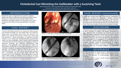
Case Description/Methods: A 90 year old female presented to the hospital for abdominal pain and unintentional weight loss. Past medical history was notable for breast cancer and colorectal cancer status post resection. Liver function tests and pancreatic markers were normal with mildly elevated alkaline phosphatase. CT abdomen and pelvis revealed a 2.8 cm dilated common bile duct (CBD), choledocholithiasis, pancreatic duct dilation and absent gallbladder. Patient however denied undergoing a cholecystectomy. Given the discrepancy of imaging and history, an abdominal ultrasound was performed demonstrating large gallstones in the gallbladder with normal CBD size. Endoscopic ultrasound (EUS) revealed a 28x26 mm mass in the ampulla. FNB was performed. The distal CBD measured 7 mm with a 35 mm proximal CBD consistent with a choledochal cyst full of large stones. Pancreatic duct measured up to 8 mm. Gallbladder was confirmed to be surgically absent. ERCP was performed which revealed an infiltrative 3-4 cm mass at the major papilla. Pancreatic and biliary sphincterotomies were performed and metal stents were placed in the ventral pancreatic duct and CBD. Biopsies taken revealed pancreatic adenocarcinoma. The following month, hepatobiliary surgery performed a Whipple procedure (pancreaticoduodenectomy). Pathology findings confirmed pancreatic adenocarcinoma with metastasis to one of twelve lymph nodes. Patient was arranged to follow up with outpatient oncology.
Discussion: Choledochal cysts can be an elusive diagnosis in elderly adults given its rarity, prevalence in children, and common presentation of vague abdominal pain. Choledochal cyst should be part of the differential diagnosis regardless of patient age especially in cases of accompanying weight loss due to its increased risk of biliary malignancy. In our case, the patient was found to have choledochal cyst misrepresented as the gallbladder and incidentally found to have pancreatic adenocarcinoma. Additionally, patients may be unreliable historians leading to misinterpretation of imaging results. Further investigation is therefore warranted in these perplexing cases for an accurate diagnosis and appropriate management.

Disclosures:
Sonia Samuel, DO, Umer Ejaz Malik, MD, Domenico Viterbo, MD. B0034 - Choledochal Cyst Mimicking the Gallbladder With a Surprising Twist, ACG 2022 Annual Scientific Meeting Abstracts. Charlotte, NC: American College of Gastroenterology.
Albany Medical Center, Albany, NY
Introduction: Choledochal cysts are congenital rarities and often a pediatric diagnosis but 20% go undetected until adulthood. Etiology remains unclear but an obstructed common bile duct (CBD) has been identified as a pathogenic feature. We present a rare case of choledochal cyst initially mimicking the gallbladder in an elderly female.
Case Description/Methods: A 90 year old female presented to the hospital for abdominal pain and unintentional weight loss. Past medical history was notable for breast cancer and colorectal cancer status post resection. Liver function tests and pancreatic markers were normal with mildly elevated alkaline phosphatase. CT abdomen and pelvis revealed a 2.8 cm dilated common bile duct (CBD), choledocholithiasis, pancreatic duct dilation and absent gallbladder. Patient however denied undergoing a cholecystectomy. Given the discrepancy of imaging and history, an abdominal ultrasound was performed demonstrating large gallstones in the gallbladder with normal CBD size. Endoscopic ultrasound (EUS) revealed a 28x26 mm mass in the ampulla. FNB was performed. The distal CBD measured 7 mm with a 35 mm proximal CBD consistent with a choledochal cyst full of large stones. Pancreatic duct measured up to 8 mm. Gallbladder was confirmed to be surgically absent. ERCP was performed which revealed an infiltrative 3-4 cm mass at the major papilla. Pancreatic and biliary sphincterotomies were performed and metal stents were placed in the ventral pancreatic duct and CBD. Biopsies taken revealed pancreatic adenocarcinoma. The following month, hepatobiliary surgery performed a Whipple procedure (pancreaticoduodenectomy). Pathology findings confirmed pancreatic adenocarcinoma with metastasis to one of twelve lymph nodes. Patient was arranged to follow up with outpatient oncology.
Discussion: Choledochal cysts can be an elusive diagnosis in elderly adults given its rarity, prevalence in children, and common presentation of vague abdominal pain. Choledochal cyst should be part of the differential diagnosis regardless of patient age especially in cases of accompanying weight loss due to its increased risk of biliary malignancy. In our case, the patient was found to have choledochal cyst misrepresented as the gallbladder and incidentally found to have pancreatic adenocarcinoma. Additionally, patients may be unreliable historians leading to misinterpretation of imaging results. Further investigation is therefore warranted in these perplexing cases for an accurate diagnosis and appropriate management.

Figure: 1. Ampullary mass 2. Pancreatic duct dilation 3. Choledochal cyst. 4 Post ERCP
Disclosures:
Sonia Samuel indicated no relevant financial relationships.
Umer Ejaz Malik indicated no relevant financial relationships.
Domenico Viterbo indicated no relevant financial relationships.
Sonia Samuel, DO, Umer Ejaz Malik, MD, Domenico Viterbo, MD. B0034 - Choledochal Cyst Mimicking the Gallbladder With a Surprising Twist, ACG 2022 Annual Scientific Meeting Abstracts. Charlotte, NC: American College of Gastroenterology.
