Back


Poster Session B - Monday Morning
Category: General Endoscopy
B0300 - Unusual Presentation of a Large Duodenal Lipoma
Monday, October 24, 2022
10:00 AM – 12:00 PM ET
Location: Crown Ballroom

Has Audio
- MB
Maria Del Mar Burgos-Torres, MD
VA Caribbean Health Care System
San Juan, PR
Presenting Author(s)
Pedro A. Rosa-Cortes, MD, Maria Del Mar Burgos-Torres, MD, Jose Martin-Ortiz, MD, FACG
VA Caribbean Health Care System, San Juan, Puerto Rico
Introduction: Duodenal lipomas are rare benign tumors that are usually asymptomatic. This is the case of a patient who presented with symptomatic anemia secondary to upper GI bleeding due to a large duodenal lipoma.
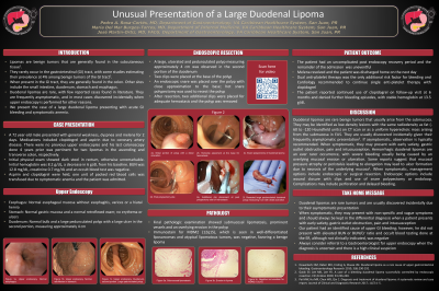
Case Description/Methods: A 72 y/o male presented with weakness, dyspnea and melena for 2 days. His last colonoscopy showed two lipomas in the ascending and transverse colon. Rectal exam showed dark stool. Hemoglobin was 8.2 g/dL. BUN was 12.8 mg/dL, creatinine 0.7 mg/dL and an occult blood test was negative. Upper endoscopy showed a large, ulcerated and pedunculated duodenal polyp in the second portion, measuring approximately 4 cm. Two hemoclips were placed at the base and hot snare polypectomy was used to remove the polyp. After removal, two hemoclips were placed for adequate hemostasis. Histopathology showed submucosal lipomatosis, prominent vessels and an overlying duodenal erosion. Immunostain for MDM2 was negative, favoring a benign lipoma. His admission was uneventful with resolution of melena. On 6-month follow-up, the patient denied further bleeding episodes, with stable hemoglobin at 13.5 g/dL.
Discussion: Lipomas are benign tumors found in the subcutaneous tissue. They rarely occur in the gastrointestinal (GI) tract, with an estimated prevalence of 4%. When present, they are generally found in the colon. Duodenal lipomas are rare, with few reported cases found in literature. Most arise from the submucosa and may be identified as a low-density lesion with the same radiodensity as fat on CT scan or as a hyperechoic mass arising from the submucosa in EUS. They are frequently asymptomatic, and in most cases discovered incidentally. If asymptomatic, observation is typically recommended. Symptoms may include early satiety, gastric outlet obstruction, pain, and intussusception. Hemorrhagic duodenal lipomas are an even rarer occurrence, with severe bleeding usually being caused by an overlying mucosal erosion or ulceration. Some reports suggest that mucosal pressure atrophy may lead to ulcer formation due to necrosis of the underlying mucosa. When symptomatic, management includes endoscopic or surgical resection. Management is dictated by operator experience, tumor size and location. Endoscopic options include wound closure with clips and use of snare polypectomy. Complications may include perforation and delayed bleeding. Duodenal lipomas may present with nonspecific and vague symptoms. For this reason, it is important to consider this in the differential diagnosis of obscure GI bleeding.

Disclosures:
Pedro A. Rosa-Cortes, MD, Maria Del Mar Burgos-Torres, MD, Jose Martin-Ortiz, MD, FACG. B0300 - Unusual Presentation of a Large Duodenal Lipoma, ACG 2022 Annual Scientific Meeting Abstracts. Charlotte, NC: American College of Gastroenterology.
VA Caribbean Health Care System, San Juan, Puerto Rico
Introduction: Duodenal lipomas are rare benign tumors that are usually asymptomatic. This is the case of a patient who presented with symptomatic anemia secondary to upper GI bleeding due to a large duodenal lipoma.
Case Description/Methods: A 72 y/o male presented with weakness, dyspnea and melena for 2 days. His last colonoscopy showed two lipomas in the ascending and transverse colon. Rectal exam showed dark stool. Hemoglobin was 8.2 g/dL. BUN was 12.8 mg/dL, creatinine 0.7 mg/dL and an occult blood test was negative. Upper endoscopy showed a large, ulcerated and pedunculated duodenal polyp in the second portion, measuring approximately 4 cm. Two hemoclips were placed at the base and hot snare polypectomy was used to remove the polyp. After removal, two hemoclips were placed for adequate hemostasis. Histopathology showed submucosal lipomatosis, prominent vessels and an overlying duodenal erosion. Immunostain for MDM2 was negative, favoring a benign lipoma. His admission was uneventful with resolution of melena. On 6-month follow-up, the patient denied further bleeding episodes, with stable hemoglobin at 13.5 g/dL.
Discussion: Lipomas are benign tumors found in the subcutaneous tissue. They rarely occur in the gastrointestinal (GI) tract, with an estimated prevalence of 4%. When present, they are generally found in the colon. Duodenal lipomas are rare, with few reported cases found in literature. Most arise from the submucosa and may be identified as a low-density lesion with the same radiodensity as fat on CT scan or as a hyperechoic mass arising from the submucosa in EUS. They are frequently asymptomatic, and in most cases discovered incidentally. If asymptomatic, observation is typically recommended. Symptoms may include early satiety, gastric outlet obstruction, pain, and intussusception. Hemorrhagic duodenal lipomas are an even rarer occurrence, with severe bleeding usually being caused by an overlying mucosal erosion or ulceration. Some reports suggest that mucosal pressure atrophy may lead to ulcer formation due to necrosis of the underlying mucosa. When symptomatic, management includes endoscopic or surgical resection. Management is dictated by operator experience, tumor size and location. Endoscopic options include wound closure with clips and use of snare polypectomy. Complications may include perforation and delayed bleeding. Duodenal lipomas may present with nonspecific and vague symptoms. For this reason, it is important to consider this in the differential diagnosis of obscure GI bleeding.

Figure: A. Snare polypectomy of duodenal lipoma
B. Post-polypectomy site
C. Resected large pedunculated duodenal polyp measuring 4 cm with distal ulceration
B. Post-polypectomy site
C. Resected large pedunculated duodenal polyp measuring 4 cm with distal ulceration
Disclosures:
Pedro Rosa-Cortes indicated no relevant financial relationships.
Maria Del Mar Burgos-Torres indicated no relevant financial relationships.
Jose Martin-Ortiz indicated no relevant financial relationships.
Pedro A. Rosa-Cortes, MD, Maria Del Mar Burgos-Torres, MD, Jose Martin-Ortiz, MD, FACG. B0300 - Unusual Presentation of a Large Duodenal Lipoma, ACG 2022 Annual Scientific Meeting Abstracts. Charlotte, NC: American College of Gastroenterology.
