Back


Poster Session A - Sunday Afternoon
Category: GI Bleeding
A0322 - Bleeding Duodenal Varix Treated With Hemostatic Clips
Sunday, October 23, 2022
5:00 PM – 7:00 PM ET
Location: Crown Ballroom

Has Audio

Joann P. Wongvravit, DO
NewYork-Presbyterian Queens Hospital
Flushing, NY
Presenting Author(s)
Joann P. Wongvravit, DO1, Ahmed Gemei, MD1, Zhi Alan Cheng, MD1, Syed Hussain, MD2
1NewYork-Presbyterian Queens Hospital, Flushing, NY; 2NewYork Presbyterian Queens Hospital, Flushing, NY
Introduction: Duodenal varices are a rare etiology of upper gastrointestinal bleeding with a prevalence of 0.4% in patients with portal hypertension undergoing esophagogastroduodenoscopy (EGD). Ectopic varices account for 1–5% of variceal bleeding, of which 17–40% are estimated to occur within the duodenum. Definitive guidelines on the management of duodenal varices, which have an estimated mortality rate of 40%, have not been established. We report the use of hemostatic clips to achieve hemostasis in a case of a bleeding duodenal varix due to portal hypertension in an orthotopic liver transplant patient with a dysfunctional mesocaval shunt.
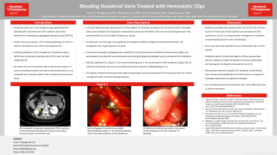
Case Description/Methods: A 42-year-old female with advanced liver cirrhosis (MELD 17) secondary to alcohol use and primary biliary cholangitis status post orthotopic liver transplantation complicated by portal vein thrombosis and biliary stricture for which she underwent mesocaval shunting and biliary reconstruction with recurrent portal hypertension. She presented with one day of bright red blood per rectum. On admission, her vital signs were significant for hypotension (81/61 mmHg) and tachycardia (132 bpm). Her hemoglobin was 7.6 g/dL (baseline 12 g/dL). Computed tomography angiography was remarkable for prominent proximal duodenal mucosal varices, portosystemic shunting with prominent splenorenal and gastric/gastroesophageal varices, and portal vein thrombosis. EGD was significant for a large ( > 5 mm) actively bleeding varix in the second portion of the duodenum, for which two hemostatic clips were successfully placed with resolution of bleeding. The patient remained hemodynamically stable post-procedure and was transferred to her transplant center for further management with no further bleeding episodes.
Discussion: Alcoholic-associated liver disease (ALD) is one of the most common causes of cirrhosis. As the incidence and prevalence of ALD continues to rise, it is important that management of duodenal variceal bleeding becomes more standardized. Only a few cases have reported the use of hemostatic clips in these patients. Treatment options include band ligation, coiling, cyanoacrylate injection, balloon-occluded retrograde transvenous obliteration, and transjugular intrahepatic portosystemic shunt. Standardized treatment modalities for duodenal variceal bleeds have not been well-established and further studies are required to investigate appropriate management strategies. Our case demonstrates that hemostatic clips can be effectively used to achieve hemostasis.

Disclosures:
Joann P. Wongvravit, DO1, Ahmed Gemei, MD1, Zhi Alan Cheng, MD1, Syed Hussain, MD2. A0322 - Bleeding Duodenal Varix Treated With Hemostatic Clips, ACG 2022 Annual Scientific Meeting Abstracts. Charlotte, NC: American College of Gastroenterology.
1NewYork-Presbyterian Queens Hospital, Flushing, NY; 2NewYork Presbyterian Queens Hospital, Flushing, NY
Introduction: Duodenal varices are a rare etiology of upper gastrointestinal bleeding with a prevalence of 0.4% in patients with portal hypertension undergoing esophagogastroduodenoscopy (EGD). Ectopic varices account for 1–5% of variceal bleeding, of which 17–40% are estimated to occur within the duodenum. Definitive guidelines on the management of duodenal varices, which have an estimated mortality rate of 40%, have not been established. We report the use of hemostatic clips to achieve hemostasis in a case of a bleeding duodenal varix due to portal hypertension in an orthotopic liver transplant patient with a dysfunctional mesocaval shunt.
Case Description/Methods: A 42-year-old female with advanced liver cirrhosis (MELD 17) secondary to alcohol use and primary biliary cholangitis status post orthotopic liver transplantation complicated by portal vein thrombosis and biliary stricture for which she underwent mesocaval shunting and biliary reconstruction with recurrent portal hypertension. She presented with one day of bright red blood per rectum. On admission, her vital signs were significant for hypotension (81/61 mmHg) and tachycardia (132 bpm). Her hemoglobin was 7.6 g/dL (baseline 12 g/dL). Computed tomography angiography was remarkable for prominent proximal duodenal mucosal varices, portosystemic shunting with prominent splenorenal and gastric/gastroesophageal varices, and portal vein thrombosis. EGD was significant for a large ( > 5 mm) actively bleeding varix in the second portion of the duodenum, for which two hemostatic clips were successfully placed with resolution of bleeding. The patient remained hemodynamically stable post-procedure and was transferred to her transplant center for further management with no further bleeding episodes.
Discussion: Alcoholic-associated liver disease (ALD) is one of the most common causes of cirrhosis. As the incidence and prevalence of ALD continues to rise, it is important that management of duodenal variceal bleeding becomes more standardized. Only a few cases have reported the use of hemostatic clips in these patients. Treatment options include band ligation, coiling, cyanoacrylate injection, balloon-occluded retrograde transvenous obliteration, and transjugular intrahepatic portosystemic shunt. Standardized treatment modalities for duodenal variceal bleeds have not been well-established and further studies are required to investigate appropriate management strategies. Our case demonstrates that hemostatic clips can be effectively used to achieve hemostasis.

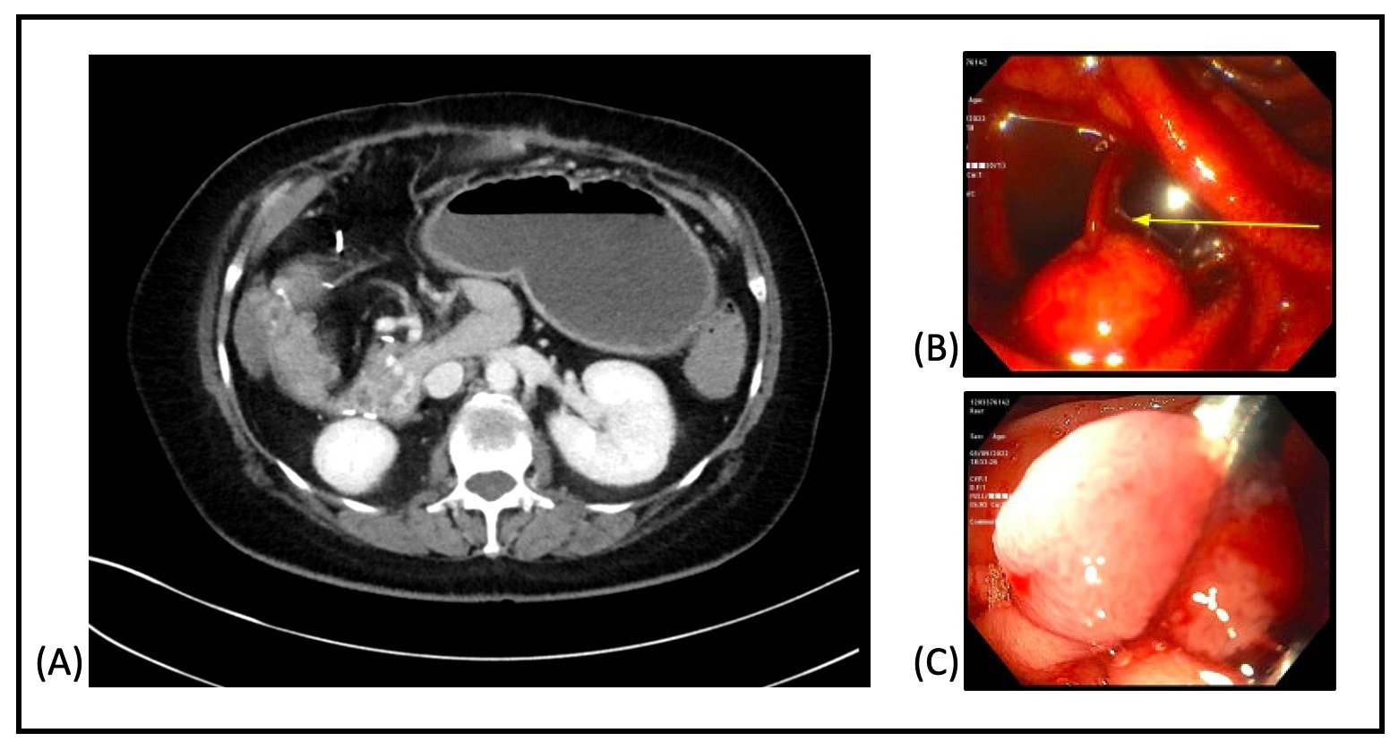
Figure: Figure 1.: (A) Computed tomography angiography (CTA) revealing prominent proximal duodenal mucosal varices, likely from the superior mesenteric vein. (B) Esophagogastroduodenoscopy (EGD) demonstrating a large (> 5 mm) actively bleeding varix in the second portion of the duodenum. (C) EGD demonstrating hemostatic clips placed on the duodenal varix with resolution of bleeding.
Disclosures:
Joann Wongvravit indicated no relevant financial relationships.
Ahmed Gemei indicated no relevant financial relationships.
Zhi Alan Cheng indicated no relevant financial relationships.
Syed Hussain indicated no relevant financial relationships.
Joann P. Wongvravit, DO1, Ahmed Gemei, MD1, Zhi Alan Cheng, MD1, Syed Hussain, MD2. A0322 - Bleeding Duodenal Varix Treated With Hemostatic Clips, ACG 2022 Annual Scientific Meeting Abstracts. Charlotte, NC: American College of Gastroenterology.
