Back


Poster Session C - Monday Afternoon
Category: Biliary/Pancreas
C0030 - Double Trouble: A Rare Anatomical Variant of the Major Papilla
Monday, October 24, 2022
3:00 PM – 5:00 PM ET
Location: Crown Ballroom

Has Audio
- NS
Naveena Sunkara, MD
Brown University
Providence, RI
Presenting Author(s)
Naveena Sunkara, MD1, Tzu-Yu Liu, MD2, Daniel Marino, MD, MBA2, Kanhai Farrakhan, MD2, Jaehoon Cho, MD3, Sarah M. Hyder, MD, MBA4
1Brown University, Providence, RI; 2Warren Alpert Medical School of Brown University, Providence, RI; 3Columbia University, New York, NY; 4Lifespan Physician Group - Brown University, East Providence, RI
Introduction: The common bile duct (CBD) and pancreatic duct (PD) join together at the level of the ampulla before opening into the duodenum via a single orifice. Biliary tree anatomic abnormalities stem from failure of rotation or recanalization during embryo development. We present a case of choledocholithaisis explored via endoscopic retrograde cholangiopancreatography (ERCP), revealing a double major papilla.
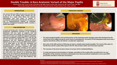
Case Description/Methods: A 61 year-old female with a history of cholecystectomy for symptomatic cholelithiasis presented to the emergency department with four weeks of epigastric pain and associated nausea and vomiting. Vitals were within normal limits and her exam was notable for epigastric tenderness. Laboratory investigation revealed WBC 11.2 x 10⁶, AST 104 IU/L, ALT 196 IU/L, total bilirubin 1.3 mg/dL, and alkaline phosphatase 433 IU/L. Abdominal ultrasound showed dilatation of the CBD to 10 mm and MRCP demonstrated a 2.9 cm filling defect in the CBD with intrahepatic and extrahepatic biliary dilation indicative of obstructing choledocholithiasis. The patient underwent ERCP which found a bulging major papilla consisting of two separate openings, both draining bile (Figures A,B). The proximal duct was deeply cannulated and contrast extended throughout the entire biliary tree. Removal of an obstructing, 23mm biliary stone was achieved by sphincterotomy and balloon extraction (Figure C). The patient was pain free and tolerated oral intake at discharge. At one week follow-up, liver function tests were improved.
Discussion: The major duodenal papilla is the terminal portion of the hepatopancreatic drainage system that develops from the ventral wall of the midgut during the fourth week of embryogenesis. By week 8-12 the hepatopancreatic ducts have a superior and inferior orifice; the inferior is usually later suppressed. Non-union of the CBD and duct of Wirsung can lead to a double major duodenal papilla. The cranial orifice opens to the bile duct and the caudal orifice opens to the PD. Only 10 other similar cases have been reported. In order to avoid unnecessary instrumentation or damage, cannulation of the caudal orifice was deferred in our case. This nonunion of the ducts has been shown to increase risk for choledocholithiasis. Recognition of this anatomic anomaly by endoscopists may reduce procedural complications such as post ERCP pancreatitis and failure of bile duct cannulation.

Disclosures:
Naveena Sunkara, MD1, Tzu-Yu Liu, MD2, Daniel Marino, MD, MBA2, Kanhai Farrakhan, MD2, Jaehoon Cho, MD3, Sarah M. Hyder, MD, MBA4. C0030 - Double Trouble: A Rare Anatomical Variant of the Major Papilla, ACG 2022 Annual Scientific Meeting Abstracts. Charlotte, NC: American College of Gastroenterology.
1Brown University, Providence, RI; 2Warren Alpert Medical School of Brown University, Providence, RI; 3Columbia University, New York, NY; 4Lifespan Physician Group - Brown University, East Providence, RI
Introduction: The common bile duct (CBD) and pancreatic duct (PD) join together at the level of the ampulla before opening into the duodenum via a single orifice. Biliary tree anatomic abnormalities stem from failure of rotation or recanalization during embryo development. We present a case of choledocholithaisis explored via endoscopic retrograde cholangiopancreatography (ERCP), revealing a double major papilla.
Case Description/Methods: A 61 year-old female with a history of cholecystectomy for symptomatic cholelithiasis presented to the emergency department with four weeks of epigastric pain and associated nausea and vomiting. Vitals were within normal limits and her exam was notable for epigastric tenderness. Laboratory investigation revealed WBC 11.2 x 10⁶, AST 104 IU/L, ALT 196 IU/L, total bilirubin 1.3 mg/dL, and alkaline phosphatase 433 IU/L. Abdominal ultrasound showed dilatation of the CBD to 10 mm and MRCP demonstrated a 2.9 cm filling defect in the CBD with intrahepatic and extrahepatic biliary dilation indicative of obstructing choledocholithiasis. The patient underwent ERCP which found a bulging major papilla consisting of two separate openings, both draining bile (Figures A,B). The proximal duct was deeply cannulated and contrast extended throughout the entire biliary tree. Removal of an obstructing, 23mm biliary stone was achieved by sphincterotomy and balloon extraction (Figure C). The patient was pain free and tolerated oral intake at discharge. At one week follow-up, liver function tests were improved.
Discussion: The major duodenal papilla is the terminal portion of the hepatopancreatic drainage system that develops from the ventral wall of the midgut during the fourth week of embryogenesis. By week 8-12 the hepatopancreatic ducts have a superior and inferior orifice; the inferior is usually later suppressed. Non-union of the CBD and duct of Wirsung can lead to a double major duodenal papilla. The cranial orifice opens to the bile duct and the caudal orifice opens to the PD. Only 10 other similar cases have been reported. In order to avoid unnecessary instrumentation or damage, cannulation of the caudal orifice was deferred in our case. This nonunion of the ducts has been shown to increase risk for choledocholithiasis. Recognition of this anatomic anomaly by endoscopists may reduce procedural complications such as post ERCP pancreatitis and failure of bile duct cannulation.

Figure: A: Two apertures of the Major Papilla: the proximal (short arrow) and distal opening to the common bile duct (long arrow) separated by 2 to 3 cm of mucosa
B: Distal opening to pancreatic duct (two short arrows)
C: Stone removal
B: Distal opening to pancreatic duct (two short arrows)
C: Stone removal
Disclosures:
Naveena Sunkara indicated no relevant financial relationships.
Tzu-Yu Liu indicated no relevant financial relationships.
Daniel Marino indicated no relevant financial relationships.
Kanhai Farrakhan indicated no relevant financial relationships.
Jaehoon Cho indicated no relevant financial relationships.
Sarah Hyder indicated no relevant financial relationships.
Naveena Sunkara, MD1, Tzu-Yu Liu, MD2, Daniel Marino, MD, MBA2, Kanhai Farrakhan, MD2, Jaehoon Cho, MD3, Sarah M. Hyder, MD, MBA4. C0030 - Double Trouble: A Rare Anatomical Variant of the Major Papilla, ACG 2022 Annual Scientific Meeting Abstracts. Charlotte, NC: American College of Gastroenterology.
