Back


Poster Session B - Monday Morning
Category: Esophagus
B0254 - A Middle-Aged Patient With an Unusual Cause of Dysphagia
Monday, October 24, 2022
10:00 AM – 12:00 PM ET
Location: Crown Ballroom

Has Audio

Prithi Choday, MD
Hemet Global Medical Center
San Jacinto, CA
Presenting Author(s)
Prithi Choday, MD1, Sabina Kumar, MS, DO2, Andrew Keldgord, DO3, Pranav Barve, MD, MPH3
1Hemet Global Medical Center, San Jacinto, CA; 2Hemet Global Medical Center, Artesia, CA; 3Hemet Global Medical Center, Hemet, CA
Introduction: Dysphagia is a common bothersome symptom affecting 3% of the US at any given time. [1] It comes with a wide range of potential causes that can be categorized into anatomic, obstructive, and neuromuscular. The most common pathophysiologic origin is from gastroesophageal reflux disease. Other causes include stroke, malignancy, and anaphylaxis. Due to the broad nature of this symptom, the rarer causes of dysphagia go unrecognized and under-diagnosed. Here we present a rare case of dysphagia in a middle-aged woman diagnosed from radiologic imaging.
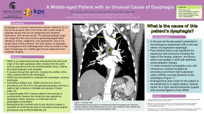
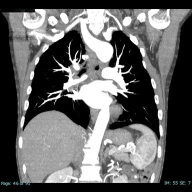
Case Description/Methods: A 65-year-old male patient presented to the emergency department with a two-day history of progressive dysphagia. Past medical history was significant for squamous cell carcinoma involving the base of the tongue, pharynx, and larynx, which was treated in 2018 with definitive chemoradiation therapy. A chest computed tomography scan with intravenous contrast revealed an anomalous prominent right subclavian artery (ARSA) coursing posterior to the esophagus (Figure 1). Arrangements were made for the patient to be transferred to a nearby tertiary medical center for a right carotid-subclavian bypass with proximal ligation of the ARSA.
Discussion: ARSA is an anatomical anomaly derived from the abnormal origin of the right subclavian artery directly from the aortic arch as opposed to from the brachiocephalic artery. It courses through the right arm, crossing the midline of the chest, passing behind the esophagus. ARSA has the potential to compress the esophagus, causing dysphagia. According to Natsis et al., ARSA was found to have a prevalence of 0.16-4.4% of the general population and has a relative high incidence in females and people of Greek origin. [2] In approximately 93% of these patient’s the anomaly is asymptomatic however the remaining may experience dysphagia, stridor, dyspnea, or chest pain due to esophageal compression. Recognizing this condition prior to any thoracic surgery is essential as unintentional injury to this artery during surgical procedures may be life-threatening. [3]

Disclosures:
Prithi Choday, MD1, Sabina Kumar, MS, DO2, Andrew Keldgord, DO3, Pranav Barve, MD, MPH3. B0254 - A Middle-Aged Patient With an Unusual Cause of Dysphagia, ACG 2022 Annual Scientific Meeting Abstracts. Charlotte, NC: American College of Gastroenterology.
1Hemet Global Medical Center, San Jacinto, CA; 2Hemet Global Medical Center, Artesia, CA; 3Hemet Global Medical Center, Hemet, CA
Introduction: Dysphagia is a common bothersome symptom affecting 3% of the US at any given time. [1] It comes with a wide range of potential causes that can be categorized into anatomic, obstructive, and neuromuscular. The most common pathophysiologic origin is from gastroesophageal reflux disease. Other causes include stroke, malignancy, and anaphylaxis. Due to the broad nature of this symptom, the rarer causes of dysphagia go unrecognized and under-diagnosed. Here we present a rare case of dysphagia in a middle-aged woman diagnosed from radiologic imaging.
Case Description/Methods: A 65-year-old male patient presented to the emergency department with a two-day history of progressive dysphagia. Past medical history was significant for squamous cell carcinoma involving the base of the tongue, pharynx, and larynx, which was treated in 2018 with definitive chemoradiation therapy. A chest computed tomography scan with intravenous contrast revealed an anomalous prominent right subclavian artery (ARSA) coursing posterior to the esophagus (Figure 1). Arrangements were made for the patient to be transferred to a nearby tertiary medical center for a right carotid-subclavian bypass with proximal ligation of the ARSA.
Discussion: ARSA is an anatomical anomaly derived from the abnormal origin of the right subclavian artery directly from the aortic arch as opposed to from the brachiocephalic artery. It courses through the right arm, crossing the midline of the chest, passing behind the esophagus. ARSA has the potential to compress the esophagus, causing dysphagia. According to Natsis et al., ARSA was found to have a prevalence of 0.16-4.4% of the general population and has a relative high incidence in females and people of Greek origin. [2] In approximately 93% of these patient’s the anomaly is asymptomatic however the remaining may experience dysphagia, stridor, dyspnea, or chest pain due to esophageal compression. Recognizing this condition prior to any thoracic surgery is essential as unintentional injury to this artery during surgical procedures may be life-threatening. [3]
- Cho et al. Prevalence and risk factors for dysphagia: a USA community study. Neurogastroenterol Motil. 2015;27(2):212-219.
- Natsis et al. The aberrant right subclavian artery: cadaveric study and literature review. Surg Radiol Anat. 2017;39(5):559-565.
- Mahmodlou et al. Aberrant Right Subclavian Artery: A Life-threatening Anomaly that should be considered during Esophagectomy. J Surg Tech Case Rep. 2014;6(2):61-63.

Figure: Coronal View
Reconstructed coronal section of computerized chest tomography with maximum intensity projection demonstrating the rise of the right subclavian artery directly from the aortic arch rather than the brachiocephalic artery.
Reconstructed coronal section of computerized chest tomography with maximum intensity projection demonstrating the rise of the right subclavian artery directly from the aortic arch rather than the brachiocephalic artery.
Disclosures:
Prithi Choday indicated no relevant financial relationships.
Sabina Kumar indicated no relevant financial relationships.
Andrew Keldgord indicated no relevant financial relationships.
Pranav Barve indicated no relevant financial relationships.
Prithi Choday, MD1, Sabina Kumar, MS, DO2, Andrew Keldgord, DO3, Pranav Barve, MD, MPH3. B0254 - A Middle-Aged Patient With an Unusual Cause of Dysphagia, ACG 2022 Annual Scientific Meeting Abstracts. Charlotte, NC: American College of Gastroenterology.
