Back


Poster Session C - Monday Afternoon
Category: Colon
C0110 - A Rare Case of Triple Synchronous Colon and Metachronous Breast Cancer in an Elderly Male
Monday, October 24, 2022
3:00 PM – 5:00 PM ET
Location: Crown Ballroom

Has Audio

Mohammed Gandam, MBBS, MD
Ascension Saint Joseph Hospital
Chicago, IL
Presenting Author(s)
Mohammed Gandam, MBBS, MD1, Aquila Fathima, MBBS, MD1, Sujatha Kailas, MD1, Alan Auerbach, MD1, George Atia, MD1, Chenyu Sun, MD1, Iqbal Ahmed, MBBS, MD2, Ramsha Faatima, MBBS3, Qurrat ul Ain Abid, MD1
1Ascension Saint Joseph Hospital, Chicago, IL; 2Loma Linda University of Health Sciences, Loma Linda, CA; 3Fathima Institute of Medical Sciences, Chicago, IL
Introduction: Although rare, Multiple Primary Malignancies (MPM) are being commonly found due to rise in elderly cancer survivor population, increased awareness and management. MPMs can be classified into synchronous (detected < 6 months) or metachronous (detected >6 months) after diagnosis of primary cancer.
Case Description/Methods: A 77-year old AA male patient with a history of gout, seizures, anemia, hypertension and hyperlipidemia initially presented with a Hb of 5.8, 8 months prior to index presentation which was treated with blood transfusion but was lost to follow up for a colonoscopy. At index presentation, his Hb was 5.5. A CT AP showed a 4 cm mass in the ascending colon, hepatosplenomegaly and a 1 cm right adrenal mass. Colonoscopy showed 4.5 cm sigmoid colon invasive moderately differentiated adenocarcinoma, a 1.5 cm hepatic flexure adenocarcinoma and a 5 cm moderately differentiated adenocarcinoma in ascending colon. CEA was noted to be 19.6. No evidence of metastasis was noted. He underwent total abdominal colectomy with 0/13 pericolonic lymphnodes without evidence of metastasis with a T3N0M0 stage. MLH-1, MSH-2, MSH-6, PMS-2 negative. A follow up CT AP for the adrenal mass and subsequent work up was suggested but he was lost to follow up. Three years later, he was found to have a 3 x 2.5 cm grade 2 invasive ductal carcinoma in the left breast with ulceration which was CDX2 and TTF1/Napsin negative, GATA-3 positive suggestive of a primary breast neoplasm. He was HER-2 negative, ER+, PR+, Ki-67 - 60.78% strong with lymphovascular and perineural invasion with a pT4 stage. A CT chest showed multiple metastatic lesions not amenable for biopsy by interventional radiology or by EBUS. He underwent left palliative mastectomy, palliative radiotherapy to spinal metastasis and palliative chemotherapy for breast cancer. CEA was 5.6.
Discussion: This patient is unique in that three large synchronous colon cancers and a large metachronous primary breast neoplasm were found in a male patient. To the best of our knowledge, this is a first reported case of a triple synchronous colon and a breast cancer in a male which is not metastatic but a metachronous cancer. Previous case reports were noted in females. Also, unique to this patient is the presence of ductal invasive carcinoma as opposed to lobular breast cancer reported previously. The association between breast and colon cancers should not be dismissed merely as metastasis and males should undergo a thorough physical exam during follow up.

Disclosures:
Mohammed Gandam, MBBS, MD1, Aquila Fathima, MBBS, MD1, Sujatha Kailas, MD1, Alan Auerbach, MD1, George Atia, MD1, Chenyu Sun, MD1, Iqbal Ahmed, MBBS, MD2, Ramsha Faatima, MBBS3, Qurrat ul Ain Abid, MD1. C0110 - A Rare Case of Triple Synchronous Colon and Metachronous Breast Cancer in an Elderly Male, ACG 2022 Annual Scientific Meeting Abstracts. Charlotte, NC: American College of Gastroenterology.
1Ascension Saint Joseph Hospital, Chicago, IL; 2Loma Linda University of Health Sciences, Loma Linda, CA; 3Fathima Institute of Medical Sciences, Chicago, IL
Introduction: Although rare, Multiple Primary Malignancies (MPM) are being commonly found due to rise in elderly cancer survivor population, increased awareness and management. MPMs can be classified into synchronous (detected < 6 months) or metachronous (detected >6 months) after diagnosis of primary cancer.
Case Description/Methods: A 77-year old AA male patient with a history of gout, seizures, anemia, hypertension and hyperlipidemia initially presented with a Hb of 5.8, 8 months prior to index presentation which was treated with blood transfusion but was lost to follow up for a colonoscopy. At index presentation, his Hb was 5.5. A CT AP showed a 4 cm mass in the ascending colon, hepatosplenomegaly and a 1 cm right adrenal mass. Colonoscopy showed 4.5 cm sigmoid colon invasive moderately differentiated adenocarcinoma, a 1.5 cm hepatic flexure adenocarcinoma and a 5 cm moderately differentiated adenocarcinoma in ascending colon. CEA was noted to be 19.6. No evidence of metastasis was noted. He underwent total abdominal colectomy with 0/13 pericolonic lymphnodes without evidence of metastasis with a T3N0M0 stage. MLH-1, MSH-2, MSH-6, PMS-2 negative. A follow up CT AP for the adrenal mass and subsequent work up was suggested but he was lost to follow up. Three years later, he was found to have a 3 x 2.5 cm grade 2 invasive ductal carcinoma in the left breast with ulceration which was CDX2 and TTF1/Napsin negative, GATA-3 positive suggestive of a primary breast neoplasm. He was HER-2 negative, ER+, PR+, Ki-67 - 60.78% strong with lymphovascular and perineural invasion with a pT4 stage. A CT chest showed multiple metastatic lesions not amenable for biopsy by interventional radiology or by EBUS. He underwent left palliative mastectomy, palliative radiotherapy to spinal metastasis and palliative chemotherapy for breast cancer. CEA was 5.6.
Discussion: This patient is unique in that three large synchronous colon cancers and a large metachronous primary breast neoplasm were found in a male patient. To the best of our knowledge, this is a first reported case of a triple synchronous colon and a breast cancer in a male which is not metastatic but a metachronous cancer. Previous case reports were noted in females. Also, unique to this patient is the presence of ductal invasive carcinoma as opposed to lobular breast cancer reported previously. The association between breast and colon cancers should not be dismissed merely as metastasis and males should undergo a thorough physical exam during follow up.

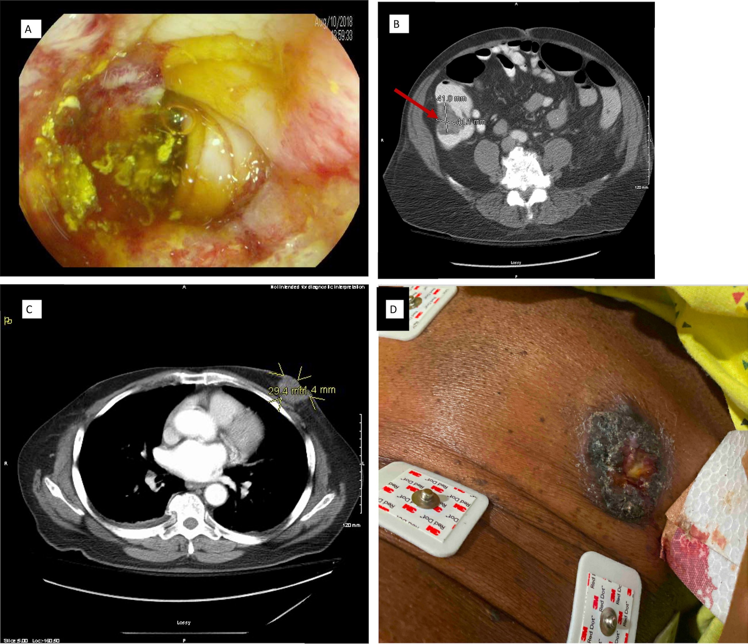
Figure: A - Sigmoid colon adenocarcinoma
B - Apple core mass in the ascending colon
C - Left breast mass without lymphadenopathy
D - Ulcerated left breast mass involving areola
B - Apple core mass in the ascending colon
C - Left breast mass without lymphadenopathy
D - Ulcerated left breast mass involving areola
Disclosures:
Mohammed Gandam indicated no relevant financial relationships.
Aquila Fathima indicated no relevant financial relationships.
Sujatha Kailas indicated no relevant financial relationships.
Alan Auerbach indicated no relevant financial relationships.
George Atia indicated no relevant financial relationships.
Chenyu Sun indicated no relevant financial relationships.
Iqbal Ahmed indicated no relevant financial relationships.
Ramsha Faatima indicated no relevant financial relationships.
Qurrat ul Ain Abid indicated no relevant financial relationships.
Mohammed Gandam, MBBS, MD1, Aquila Fathima, MBBS, MD1, Sujatha Kailas, MD1, Alan Auerbach, MD1, George Atia, MD1, Chenyu Sun, MD1, Iqbal Ahmed, MBBS, MD2, Ramsha Faatima, MBBS3, Qurrat ul Ain Abid, MD1. C0110 - A Rare Case of Triple Synchronous Colon and Metachronous Breast Cancer in an Elderly Male, ACG 2022 Annual Scientific Meeting Abstracts. Charlotte, NC: American College of Gastroenterology.
