Back


Poster Session C - Monday Afternoon
Category: Colon
C0112 - Osseous Metaplasia in a Colon Polyp
Monday, October 24, 2022
3:00 PM – 5:00 PM ET
Location: Crown Ballroom

Has Audio

Parth M. Patel, MD
Henry Ford Jackson
Jackson, MI
Presenting Author(s)
Parth M. Patel, MD, Hassan Zreik, MD, Harjinder Singh, MD, Sruthi Ramanan, MD, Shamik Parikh, MD, Vivek Kak, MD, Vishal Gupta, MD, PhD
Henry Ford Jackson, Jackson, MI
Introduction: Osseous metaplasia is a heterotopic bone formation that has been described in a wide variety of tissue types, both neoplastic and inflammatory lesions. However, its occurrence in colorectal cancer is exceedingly rare. Bone formation in a rectal polyp was first described in 1981. We describe a rare case of osseous metaplasia in rectal mucosa with a nearby hyperplastic polyp.
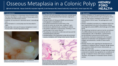
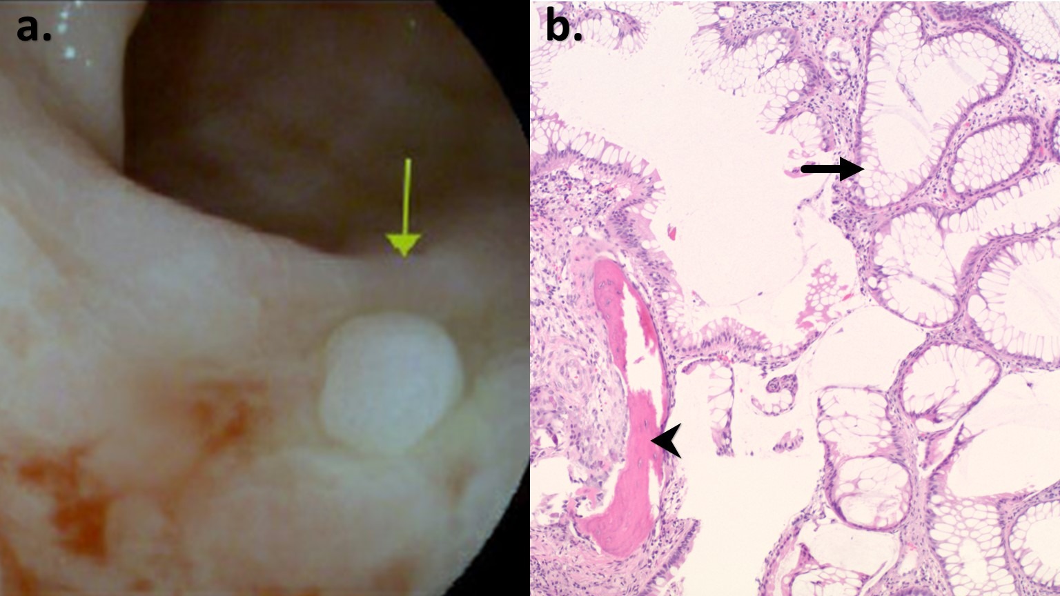
Case Description/Methods: A 36-year-old male presented to the clinic complaining of diarrhea and hematochezia that had been ongoing for several years. He had a history of infrequent NSAID use but denied anticoagulant or antiplatelet use. He noticed hematochezia several times a day. He did not notice any rectal mass, swelling, or pain. He denied any abdominal pain, vomiting, or melena. The patient underwent a colonoscopy, revealing a 7 mm sessile polyp in the rectum (Figure 1a). The polyp was removed by a cold snare and was sent for histopathological evaluation. Histopathology of the hyperplastic polyp demonstrated osseous metaplasia with foreign body reaction (Figure 1b).
Discussion: Osseous metaplasia of the gastrointestinal tract is an exceedingly rare phenomenon with a prevalence of less than 1%, with osseous metaplasia of the rectum accounting for 0.4% of cases. Although osseous metaplasia is a well-defined phenomenon, the pathogenesis is still unclear. Osseous metaplasia secondary to a malignant lesion has been hypothesized to be due to the presence of bone morphogenetic protein (BMP), which belongs to the TGFβ family.
The underlying mechanism seems to be the recruitment of undifferentiated stromal mesenchymal cells into osteoprogenitor cells or fibroblasts, a phenomenon termed epithelial-mesenchymal transformation. Kypson et al. demonstrated the overexpression of BMP-2 in tumor cells from cases of rectal adenocarcinoma with osseous metaplasia compared to those lacking bone formation. An alternative mechanism proposed is the increased expression of osteocalcin and upregulation of type-1 collagen and osteonectin, markers of bone matrix synthesis. Multiple theories exist regarding the development of metaplasia in malignant lesions. However, benign lesions have only been linked with chronic inflammation, as described in our case.
Given the rarity of this condition, no large trials have been conducted to establish clear guidelines on surveillance of benign or malignant masses with osseous metaplasia. Therefore, the prognosis in metastatic lesions remains unclear.

Disclosures:
Parth M. Patel, MD, Hassan Zreik, MD, Harjinder Singh, MD, Sruthi Ramanan, MD, Shamik Parikh, MD, Vivek Kak, MD, Vishal Gupta, MD, PhD. C0112 - Osseous Metaplasia in a Colon Polyp, ACG 2022 Annual Scientific Meeting Abstracts. Charlotte, NC: American College of Gastroenterology.
Henry Ford Jackson, Jackson, MI
Introduction: Osseous metaplasia is a heterotopic bone formation that has been described in a wide variety of tissue types, both neoplastic and inflammatory lesions. However, its occurrence in colorectal cancer is exceedingly rare. Bone formation in a rectal polyp was first described in 1981. We describe a rare case of osseous metaplasia in rectal mucosa with a nearby hyperplastic polyp.
Case Description/Methods: A 36-year-old male presented to the clinic complaining of diarrhea and hematochezia that had been ongoing for several years. He had a history of infrequent NSAID use but denied anticoagulant or antiplatelet use. He noticed hematochezia several times a day. He did not notice any rectal mass, swelling, or pain. He denied any abdominal pain, vomiting, or melena. The patient underwent a colonoscopy, revealing a 7 mm sessile polyp in the rectum (Figure 1a). The polyp was removed by a cold snare and was sent for histopathological evaluation. Histopathology of the hyperplastic polyp demonstrated osseous metaplasia with foreign body reaction (Figure 1b).
Discussion: Osseous metaplasia of the gastrointestinal tract is an exceedingly rare phenomenon with a prevalence of less than 1%, with osseous metaplasia of the rectum accounting for 0.4% of cases. Although osseous metaplasia is a well-defined phenomenon, the pathogenesis is still unclear. Osseous metaplasia secondary to a malignant lesion has been hypothesized to be due to the presence of bone morphogenetic protein (BMP), which belongs to the TGFβ family.
The underlying mechanism seems to be the recruitment of undifferentiated stromal mesenchymal cells into osteoprogenitor cells or fibroblasts, a phenomenon termed epithelial-mesenchymal transformation. Kypson et al. demonstrated the overexpression of BMP-2 in tumor cells from cases of rectal adenocarcinoma with osseous metaplasia compared to those lacking bone formation. An alternative mechanism proposed is the increased expression of osteocalcin and upregulation of type-1 collagen and osteonectin, markers of bone matrix synthesis. Multiple theories exist regarding the development of metaplasia in malignant lesions. However, benign lesions have only been linked with chronic inflammation, as described in our case.
Given the rarity of this condition, no large trials have been conducted to establish clear guidelines on surveillance of benign or malignant masses with osseous metaplasia. Therefore, the prognosis in metastatic lesions remains unclear.

Figure: Figure 1: a. 7 mm rectal polyp (yellow arrow) seen on colonoscopy and subsequently removed by cold snare technique. b. Histology specimen (x80 magnification) obtained from a rectal mucosa biopsy demonstrating osseous metaplasia (Arrowhead) with benign foreign body reaction. Figure also demonstrates the osseous metaplasia surrounded by normal colonic mucosa and crypts (Arrow).
Disclosures:
Parth Patel indicated no relevant financial relationships.
Hassan Zreik indicated no relevant financial relationships.
Harjinder Singh indicated no relevant financial relationships.
Sruthi Ramanan indicated no relevant financial relationships.
Shamik Parikh indicated no relevant financial relationships.
Vivek Kak indicated no relevant financial relationships.
Vishal Gupta indicated no relevant financial relationships.
Parth M. Patel, MD, Hassan Zreik, MD, Harjinder Singh, MD, Sruthi Ramanan, MD, Shamik Parikh, MD, Vivek Kak, MD, Vishal Gupta, MD, PhD. C0112 - Osseous Metaplasia in a Colon Polyp, ACG 2022 Annual Scientific Meeting Abstracts. Charlotte, NC: American College of Gastroenterology.
