Back


Poster Session C - Monday Afternoon
Category: Colon
C0155 - Colonic Carcinoma With Neuroendocrine Features Diagnosed in the Setting of Enterococcus faecalis Bacteremia, Endocarditis and Embolic Phenomenon
Monday, October 24, 2022
3:00 PM – 5:00 PM ET
Location: Crown Ballroom

Has Audio

Gassan Kassim, MD
Mayo Clinic
Scottsdale, AZ
Presenting Author(s)
Gassan Kassim, MD1, Allon Kahn, MD1, Dora Lam-Himlin, MD1, Christine Menias, MD1, Tisha Lundsford, MD2
1Mayo Clinic, Scottsdale, AZ; 2Mayo Clinic Arizona, Scottsdale, AZ
Introduction: A 77-year-old man with a history of hypertension, hyperlipidemia, prior ischemic stroke, and chronic back pain presented with fever and confusion 3 days after undergoing a lumbar epidural steroid injection.
Case Description/Methods: A 77-year-old man with a history of hypertension, hyperlipidemia, prior ischemic stroke, and chronic back pain presented with fever and confusion 3 days after undergoing a lumbar epidural steroid injection.
Multiple sets of blood cultures grew Enterococcus faecalis. His CRP was elevated at 86. An MRI scan of the spine was negative for epidural abscess. Brain MRI revealed multiple acute embolic infarcts. Transthoracic echocardiography revealed a mobile 8mm vegetation, and transesophageal echocardiography confirmed mitral valve endocarditis. CT of the abdomen and pelvis revealed colonic wall thickening at the hepatic flexure with coexistent fat stranding and lymphadenopathy.
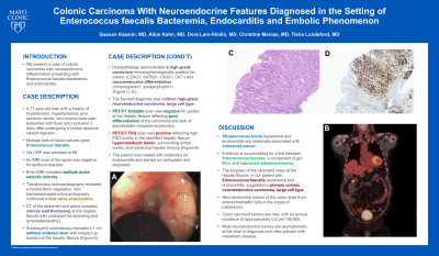
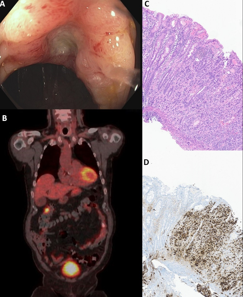
Subsequent colonoscopy revealed a 1 cm solitary cratered ulcer with heaped up borders at the hepatic flexure (Figure 1A). Histopathology demonstrated a high-grade carcinoma immunophenotypically positive for colonic (CDX2+, SATB2+, CK20+, CK7-) and neuroendocrine differentiation (chromogranin+, synaptophysin+) (Figure 2C, 2D). The favored diagnosis was colonic high-grade neuroendocrine carcinoma, large cell type. PET/CT Dotatate scan was negative for uptake at the hepatic flexure reflecting poor differentiation of the carcinoma and lack of somatostatin receptors expression. Subsequent PET/CT FDG scan was positive reflecting high FDG avidity in the identified hepatic flexure hypermetabolic lesion, surrounding lymph nodes, and numerous hepatic lesions (Figure 1B).
The patient was treated with antibiotics for endocarditis and started on carboplatin and etoposide.
Discussion: Classically, Streptococcus bovis bacteremia and endocarditis are associated with colorectal cancer. Evidence is accumulating for a link between Enterococcus faecalis, a component of gut flora, and colorectal adenocarcinoma. The biopsies of the ulcerated mass at the hepatic flexure, in our patient with Enterococcus faecalis bacteremia and endocarditis, suggested a primary colonic neuroendocrine carcinoma, large cell type. Neuroendocrine tumors of the colon arise from enterochromaffin cells in the crypts of Lieberkuhn. Colon carcinoid tumors are rare, with an annual incidence of approximately 0.2 per 100,000. Most neuroendocrine tumors are asymptomatic at the time of diagnosis and often present with metastatic disease.

Disclosures:
Gassan Kassim, MD1, Allon Kahn, MD1, Dora Lam-Himlin, MD1, Christine Menias, MD1, Tisha Lundsford, MD2. C0155 - Colonic Carcinoma With Neuroendocrine Features Diagnosed in the Setting of Enterococcus faecalis Bacteremia, Endocarditis and Embolic Phenomenon, ACG 2022 Annual Scientific Meeting Abstracts. Charlotte, NC: American College of Gastroenterology.
1Mayo Clinic, Scottsdale, AZ; 2Mayo Clinic Arizona, Scottsdale, AZ
Introduction: A 77-year-old man with a history of hypertension, hyperlipidemia, prior ischemic stroke, and chronic back pain presented with fever and confusion 3 days after undergoing a lumbar epidural steroid injection.
Case Description/Methods: A 77-year-old man with a history of hypertension, hyperlipidemia, prior ischemic stroke, and chronic back pain presented with fever and confusion 3 days after undergoing a lumbar epidural steroid injection.
Multiple sets of blood cultures grew Enterococcus faecalis. His CRP was elevated at 86. An MRI scan of the spine was negative for epidural abscess. Brain MRI revealed multiple acute embolic infarcts. Transthoracic echocardiography revealed a mobile 8mm vegetation, and transesophageal echocardiography confirmed mitral valve endocarditis. CT of the abdomen and pelvis revealed colonic wall thickening at the hepatic flexure with coexistent fat stranding and lymphadenopathy.
Subsequent colonoscopy revealed a 1 cm solitary cratered ulcer with heaped up borders at the hepatic flexure (Figure 1A). Histopathology demonstrated a high-grade carcinoma immunophenotypically positive for colonic (CDX2+, SATB2+, CK20+, CK7-) and neuroendocrine differentiation (chromogranin+, synaptophysin+) (Figure 2C, 2D). The favored diagnosis was colonic high-grade neuroendocrine carcinoma, large cell type. PET/CT Dotatate scan was negative for uptake at the hepatic flexure reflecting poor differentiation of the carcinoma and lack of somatostatin receptors expression. Subsequent PET/CT FDG scan was positive reflecting high FDG avidity in the identified hepatic flexure hypermetabolic lesion, surrounding lymph nodes, and numerous hepatic lesions (Figure 1B).
The patient was treated with antibiotics for endocarditis and started on carboplatin and etoposide.
Discussion: Classically, Streptococcus bovis bacteremia and endocarditis are associated with colorectal cancer. Evidence is accumulating for a link between Enterococcus faecalis, a component of gut flora, and colorectal adenocarcinoma. The biopsies of the ulcerated mass at the hepatic flexure, in our patient with Enterococcus faecalis bacteremia and endocarditis, suggested a primary colonic neuroendocrine carcinoma, large cell type. Neuroendocrine tumors of the colon arise from enterochromaffin cells in the crypts of Lieberkuhn. Colon carcinoid tumors are rare, with an annual incidence of approximately 0.2 per 100,000. Most neuroendocrine tumors are asymptomatic at the time of diagnosis and often present with metastatic disease.

Figure: Figure 1A: Colonoscopy revealing a 1 cm solitary cratered ulcer at the hepatic flexure.
Figure 1B: PET/CT FDG scan showing high FDG avidity at hepatic flexure.
Figure 1C: Histopathology H&E stain showing poorly differentiated carcinoma.
Figure 1D: Histopathology chromogranin stain positivity confirming neuroendocrine differentiation
Figure 1B: PET/CT FDG scan showing high FDG avidity at hepatic flexure.
Figure 1C: Histopathology H&E stain showing poorly differentiated carcinoma.
Figure 1D: Histopathology chromogranin stain positivity confirming neuroendocrine differentiation
Disclosures:
Gassan Kassim indicated no relevant financial relationships.
Allon Kahn: MiMedx – Consultant.
Dora Lam-Himlin indicated no relevant financial relationships.
Christine Menias indicated no relevant financial relationships.
Tisha Lundsford indicated no relevant financial relationships.
Gassan Kassim, MD1, Allon Kahn, MD1, Dora Lam-Himlin, MD1, Christine Menias, MD1, Tisha Lundsford, MD2. C0155 - Colonic Carcinoma With Neuroendocrine Features Diagnosed in the Setting of Enterococcus faecalis Bacteremia, Endocarditis and Embolic Phenomenon, ACG 2022 Annual Scientific Meeting Abstracts. Charlotte, NC: American College of Gastroenterology.
