Back


Poster Session B - Monday Morning
Category: GI Bleeding
B0317 - Treatment of Cardiofundal Gastric Varices From Splenic Vein Occlusion: A Different Phenotype and Framework for Treatment?
Monday, October 24, 2022
10:00 AM – 12:00 PM ET
Location: Crown Ballroom

Has Audio

Thomas Wang, MD
Brigham and Women's Hospital
Boston, MA
Presenting Author(s)
Thomas Wang, MD1, Kanwal Bains, MBBS2, Marvin Ryou, MD3
1Brigham and Women's Hospital, Boston, MA; 2Brigham and Women’s Hospital, Boston, MA; 3Brigham & Women's Hospital, Boston, MA
Introduction: Cardiofundal gastric varices can result from left-sided portal hypertension due to isolated splenic vein occlusion, either from intraluminal thrombosis or extrinsic compression of the splenic vein. These patients often do not have systemic portal hypertension or cirrhosis. Treatment of non-cirrhosis related cardiofundal varices can be challenging, as conventional gastric variceal therapies such as transjugular intrahepatic portosystemic shunt (TIPS) or balloon occluded retrograde transvenous obliteration (BRTO) are ineffective for left sided portal hypertension. We aimed to describe our center’s experience and approach in treatment of cardiofundal varices in the setting of isolated splenic vein occlusion.
Methods: A retrospective review of all patients who presented with concern for bleeding from cardiofundal varices at two tertiary hospitals was performed from Jan 2018 to May 2022. Patients were included if they received treatment for cardiofundal varices from isolated splenic vein occlusion, and excluded if they had been previously diagnosed with cirrhosis or portal vein thrombosis.
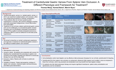
Results: We found a total of 8 patients who met the study’s inclusion and exclusion criteria. 7/8 (87.5%) were male and the mean age was 58. Etiology of splenic vein occlusion included pancreatic adenocarcinoma (n=4), necrotizing pancreatitis (n=2), and carcinoid/neuroendocrine tumors (n=2); see Table 1. Maximum variceal cross section diameter ranged from 4-10mm, and were often small in size and diffuse. All patients underwent initial endoscopic therapy, which included EUS guided coil embolization (n=6) and rubber band ligation (n=3), with 1 patient receiving both. Coil embolization was performed as standalone (n=1), with Gelfoam (n=4), or with cyanoacrylate glue (n=1). Technical success and ability to achieve hemostasis on index procedure were both 100%. 4 patients required re-intervention, with 2 refractory to endoscopic therapy requiring splenic artery embolization and splenectomy.
Discussion: Coil embolization and/or band ligation can be effective initial endoscopic therapies for non cirrhotic cardiofundal variceal bleeding. Cardiofundal varices from splenic vein occlusion on EUS often appear much smaller in size as compared to those from cirrhosis, which may preclude coil embolization – in these cases, band ligation may be a more preferable option. For patients who fail initial endoscopic therapy and require significant transfusions, splenic artery embolization and splenectomy can be considered.
Disclosures:
Thomas Wang, MD1, Kanwal Bains, MBBS2, Marvin Ryou, MD3. B0317 - Treatment of Cardiofundal Gastric Varices From Splenic Vein Occlusion: A Different Phenotype and Framework for Treatment?, ACG 2022 Annual Scientific Meeting Abstracts. Charlotte, NC: American College of Gastroenterology.
1Brigham and Women's Hospital, Boston, MA; 2Brigham and Women’s Hospital, Boston, MA; 3Brigham & Women's Hospital, Boston, MA
Introduction: Cardiofundal gastric varices can result from left-sided portal hypertension due to isolated splenic vein occlusion, either from intraluminal thrombosis or extrinsic compression of the splenic vein. These patients often do not have systemic portal hypertension or cirrhosis. Treatment of non-cirrhosis related cardiofundal varices can be challenging, as conventional gastric variceal therapies such as transjugular intrahepatic portosystemic shunt (TIPS) or balloon occluded retrograde transvenous obliteration (BRTO) are ineffective for left sided portal hypertension. We aimed to describe our center’s experience and approach in treatment of cardiofundal varices in the setting of isolated splenic vein occlusion.
Methods: A retrospective review of all patients who presented with concern for bleeding from cardiofundal varices at two tertiary hospitals was performed from Jan 2018 to May 2022. Patients were included if they received treatment for cardiofundal varices from isolated splenic vein occlusion, and excluded if they had been previously diagnosed with cirrhosis or portal vein thrombosis.
Results: We found a total of 8 patients who met the study’s inclusion and exclusion criteria. 7/8 (87.5%) were male and the mean age was 58. Etiology of splenic vein occlusion included pancreatic adenocarcinoma (n=4), necrotizing pancreatitis (n=2), and carcinoid/neuroendocrine tumors (n=2); see Table 1. Maximum variceal cross section diameter ranged from 4-10mm, and were often small in size and diffuse. All patients underwent initial endoscopic therapy, which included EUS guided coil embolization (n=6) and rubber band ligation (n=3), with 1 patient receiving both. Coil embolization was performed as standalone (n=1), with Gelfoam (n=4), or with cyanoacrylate glue (n=1). Technical success and ability to achieve hemostasis on index procedure were both 100%. 4 patients required re-intervention, with 2 refractory to endoscopic therapy requiring splenic artery embolization and splenectomy.
Discussion: Coil embolization and/or band ligation can be effective initial endoscopic therapies for non cirrhotic cardiofundal variceal bleeding. Cardiofundal varices from splenic vein occlusion on EUS often appear much smaller in size as compared to those from cirrhosis, which may preclude coil embolization – in these cases, band ligation may be a more preferable option. For patients who fail initial endoscopic therapy and require significant transfusions, splenic artery embolization and splenectomy can be considered.
| Case | Etiology of Splenic Vein Thrombosis | Maximum variceal cross section diameter (mm) | Treatment on Index Procedure | Follow up |
| 1 | Pancreatic adenocarcinoma | 5 | Coil embolization + Gelfoam | Repeat EGD with deflation of IGV1 |
| 2 | Pancreatic adenocarcinoma | 4 | Band ligation | Repeat EGD pending |
| 3 | Metastatic carcinoid tumor | 5 | Coil embolization | One additional EUS guided coil embolization in 2 weeks due to bleeding. Bleeding persisted, so referred to IR. BRTO attempted but no shunt, so ultimately underwent splenectomy. |
| 4 | Necrotizing pancreatitis | 4.5 | Coil embolization + Gelfoam | No recurrent bleeding |
| 5 | Pancreatic adenocarcinoma | 4 | Band ligation | Two additional endoscopic sessions performed for bleeding, first with coil embolization + Gelfoam, second with repeat banding (all within 2-3 months). |
| 6 | Pancreatic adenocarcinoma | 10 | Coil embolization + cyanoacrylate glue | Presented with rebleeding from IGV1 within 3 months, received Hemospray followed by splenic artery embolization. |
| 7 | Necrotizing pancreatitis | 4 | Coil embolization + Gelfoam | Repeat intervention in 3 weeks with coil embolization + Gelfoam + banding. No recurrent bleeding, IGV1 improved. |
| 8 | Pancreatic neuroendocrine tumor | 8 | Coil embolization + Gelfoam + band ligation | Repeat intervention in 2 months with repeat coil embolization + Gelfoam + banding. |
Table: Individual patient description on etiology of splenic vein thrombosis, treatment on index procedure, and follow up
Disclosures:
Thomas Wang indicated no relevant financial relationships.
Kanwal Bains indicated no relevant financial relationships.
Marvin Ryou: Boston Scientific – Consultant. Cook – Consultant. EnteraSense – Consultant, Owner/Ownership Interest. Fuji – Consultant. GI Windows – Consultant, Owner/Ownership Interest. Olympus – Consultant.
Thomas Wang, MD1, Kanwal Bains, MBBS2, Marvin Ryou, MD3. B0317 - Treatment of Cardiofundal Gastric Varices From Splenic Vein Occlusion: A Different Phenotype and Framework for Treatment?, ACG 2022 Annual Scientific Meeting Abstracts. Charlotte, NC: American College of Gastroenterology.

