Back


Poster Session B - Monday Morning
Category: Esophagus
B0259 - Esophageal Mucosal Calcinosis: A Rare Site of Calcium Deposition in End-Stage Renal Disease
Monday, October 24, 2022
10:00 AM – 12:00 PM ET
Location: Crown Ballroom

Has Audio
.jpg)
Jeffrey S. Baek
Parkside Medical Group
Rancho Cucamonga, CA
Presenting Author(s)
Jeffrey S. Baek, 1, Xiaoyan Zhou, MD2, Hussein Abidali, DO3
1Parkside Medical Group, Upland, CA; 2San Antonio Regional Hospital, Upland, CA; 3Digestive Health Inc., Rancho Cucamonga, CA
Introduction: Calciphylaxis, otherwise known as calcium uremic arteriolopathy, is defined as calcium deposition around blood vessels in skin and fat tissue which occurs in 1-4% of patients with end-stage renal disease (ESRD). Calcium deposition in the esophagus is extremely rare; to date, there have been only four cases reported worldwide.
We report the fifth case of esophageal mucosal calcinosis occurring in a young male with ESRD.
Case Description/Methods: A 37-year-old Thai man with ESRD on peritoneal dialysis since 2005 presented with generalized weakness and odynophagia due to oral ulcers, resulting in poor PO intake. He denied drinking alcohol, illicit drug use, or smoking.
On exam his abdomen was soft, non-distended, non-tender, without any guarding.
Past medical history included hypertension and COVID-19 in January 2022.
Laboratory tests revealed neutropenia and pancytopenia, hyperphosphatemia, and hypocalcemia. EGD revealed distal esophageal esophagitis and hemorrhagic erosive gastropathy. Biopsy showed ulcerative esophagitis with dystrophic calcification, consistent with esophageal mucosal calcinosis .No intestinal metaplasia was noted. Immunohistochemistry was negative for CMV, HSV1, and HSV2.
The patient was treated with pantoprazole 40mg IV every 12 hours, Magic Mouthwash 5ml qid, and Carafate 10mg qid.
He was transferred to a cancer center where he had a bone marrow biopsy formed which was negative. His symptoms resolved and the patient was discharged to home.
Discussion: Esophageal mucosal calcinosis is extremely rare. It is due to a combination of factors involving acidosis and the phenotypic differentiation (and apoptosis) of vascular smooth muscle cells (VSMC) into chondrocytes or osteoblast-like cells. These changes, along with the passive accumulation of calcium and phosphate, induce calcification. Acidosis is well-known to promote inflammation of the arterial walls, releasing cytokines that induce vascular calcification.
The benefits of treatment with sodium thiosulfate remain unclear. An ample collection of cases should help devise standardized treatment options and establish management guidelines for this condition.

Disclosures:
Jeffrey S. Baek, 1, Xiaoyan Zhou, MD2, Hussein Abidali, DO3. B0259 - Esophageal Mucosal Calcinosis: A Rare Site of Calcium Deposition in End-Stage Renal Disease, ACG 2022 Annual Scientific Meeting Abstracts. Charlotte, NC: American College of Gastroenterology.
1Parkside Medical Group, Upland, CA; 2San Antonio Regional Hospital, Upland, CA; 3Digestive Health Inc., Rancho Cucamonga, CA
Introduction: Calciphylaxis, otherwise known as calcium uremic arteriolopathy, is defined as calcium deposition around blood vessels in skin and fat tissue which occurs in 1-4% of patients with end-stage renal disease (ESRD). Calcium deposition in the esophagus is extremely rare; to date, there have been only four cases reported worldwide.
We report the fifth case of esophageal mucosal calcinosis occurring in a young male with ESRD.
Case Description/Methods: A 37-year-old Thai man with ESRD on peritoneal dialysis since 2005 presented with generalized weakness and odynophagia due to oral ulcers, resulting in poor PO intake. He denied drinking alcohol, illicit drug use, or smoking.
On exam his abdomen was soft, non-distended, non-tender, without any guarding.
Past medical history included hypertension and COVID-19 in January 2022.
Laboratory tests revealed neutropenia and pancytopenia, hyperphosphatemia, and hypocalcemia. EGD revealed distal esophageal esophagitis and hemorrhagic erosive gastropathy. Biopsy showed ulcerative esophagitis with dystrophic calcification, consistent with esophageal mucosal calcinosis .No intestinal metaplasia was noted. Immunohistochemistry was negative for CMV, HSV1, and HSV2.
The patient was treated with pantoprazole 40mg IV every 12 hours, Magic Mouthwash 5ml qid, and Carafate 10mg qid.
He was transferred to a cancer center where he had a bone marrow biopsy formed which was negative. His symptoms resolved and the patient was discharged to home.
Discussion: Esophageal mucosal calcinosis is extremely rare. It is due to a combination of factors involving acidosis and the phenotypic differentiation (and apoptosis) of vascular smooth muscle cells (VSMC) into chondrocytes or osteoblast-like cells. These changes, along with the passive accumulation of calcium and phosphate, induce calcification. Acidosis is well-known to promote inflammation of the arterial walls, releasing cytokines that induce vascular calcification.
The benefits of treatment with sodium thiosulfate remain unclear. An ample collection of cases should help devise standardized treatment options and establish management guidelines for this condition.

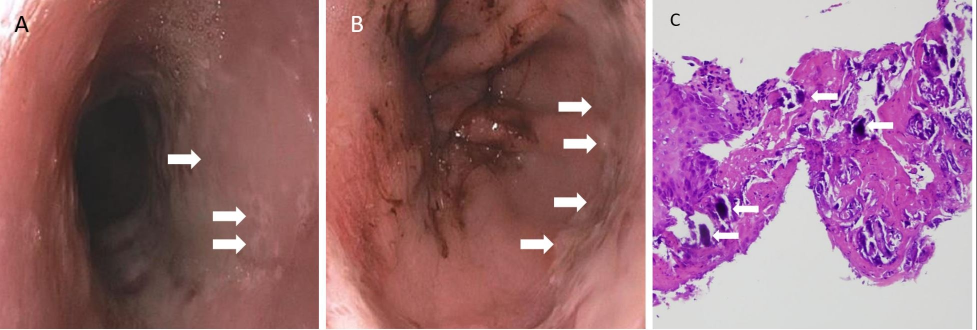
Figure: A. Circumferential thick white esophageal plaques with friability and circular erosions along the mid esophagus spanning down to the GE junction.
B. Circumferential thick white esophageal plaques with friability and circular erosions along the GE junction. Blood seen in the center is due to severe hemorrhagic gastropathy from coagulopathy.
C. Biopsy showing ulcerated esophagitis with basophilic calcium deposits(white arrows) within the fibrinopurulent exudate and beneath the squamous mucosa (H&E, original magnification 200x).
Disclosures:
Jeffrey Baek indicated no relevant financial relationships.
Xiaoyan Zhou indicated no relevant financial relationships.
Hussein Abidali indicated no relevant financial relationships.
Jeffrey S. Baek, 1, Xiaoyan Zhou, MD2, Hussein Abidali, DO3. B0259 - Esophageal Mucosal Calcinosis: A Rare Site of Calcium Deposition in End-Stage Renal Disease, ACG 2022 Annual Scientific Meeting Abstracts. Charlotte, NC: American College of Gastroenterology.

