Back


Poster Session C - Monday Afternoon
Category: GI Bleeding
C0345 - Brunner’s Gland Hamartoma: A Non-Malignant Unusual Cause of Upper Gastrointestinal Bleeding in a 77-Year-Old Man
Monday, October 24, 2022
3:00 PM – 5:00 PM ET
Location: Crown Ballroom

Has Audio

Busara Songtanin, MD
Texas Tech University Health Sciences Center
Lubbock, Texas
Presenting Author(s)
Busara Songtanin, MD1, Daoud Arif, MD1, Kanak Das, MD2
1Texas Tech University Health Sciences Center, Lubbock, TX; 2Texas Tech University Health Sciences Center and University Medical Center, Lubbock, TX
Introduction: Brunner’s gland hamartoma is a rare benign duodenal tumor. It is usually found incidentally on the esophagogastroduodenoscopy (EGD), but some patients present with gastrointestinal bleeding.
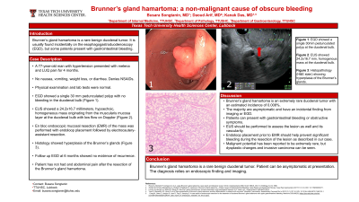
Case Description/Methods: A 77-year-old man with past medical history of hypertension presented to the hospital with melena and pain in the left upper quadrant for four months. The pain was sharp and radiated to the back. He denied nausea, vomiting, weight loss, and diarrhea. Patient had not taken NSAIDs. Vital signs were normal. Abdominal examination was soft, nontender, and without a palpable mass. Laboratory included Hb 15.2 g/dL and Hct 46.8%. EGD showed a single 30 mm pedunculated polyp with no bleeding in the duodenal bulb. Endoscopic ultrasound (EUS) showed a 24.2x16.7 millimeters, hypoechoic, homogeneous mass originating from the muscularis mucosa layer at the duodenal bulb with low flow on Doppler. En bloc endoscopic mucosal resection (EMR) of the mass was performed with the help of endoloop placement followed by electrocautery-assistant resection and endoclip placement to close the resected area. The patient tolerated the procedure well without any consequence. Histopathology of the mass showed hyperplasia of Brunner's glands. Follow up endoscopy at 6 month revealed healed scar at the resected site without any endoscopic evidence of recurrence.
Discussion: Brunner’s gland hamartoma is an extremely rare duodenal tumor with an estimated incidence of 0.008%. The name arises from the Brunner’s glands which are acinotubular glands that secrete alkaline fluid. The majority of patients are asymptomatic and have an incidental finding from the imaging or EGD. Patients commonly present with gastrointestinal bleeding and obstructive symptoms. EGD finding usually reveal a pedunculated mass 1-2cm in size located at duodenal bulb area consistent with Brunner’s gland distribution. The diagnosis relies on endoscopic finding and imaging. The pathology of the tissue yields the definitive diagnosis. Often this polyp develops into thick wide stalk which may contains large blood vessel; EUS should be performed to assess the lesion as well and its vascularity. Endoloop placement prior to EMR should help prevent significant bleeding during the resection of the lesion as described in our case. Malignant potential has been reported to be extremely rare, but dysplastic changes and invasive carcinoma can be seen.

Disclosures:
Busara Songtanin, MD1, Daoud Arif, MD1, Kanak Das, MD2. C0345 - Brunner’s Gland Hamartoma: A Non-Malignant Unusual Cause of Upper Gastrointestinal Bleeding in a 77-Year-Old Man, ACG 2022 Annual Scientific Meeting Abstracts. Charlotte, NC: American College of Gastroenterology.
1Texas Tech University Health Sciences Center, Lubbock, TX; 2Texas Tech University Health Sciences Center and University Medical Center, Lubbock, TX
Introduction: Brunner’s gland hamartoma is a rare benign duodenal tumor. It is usually found incidentally on the esophagogastroduodenoscopy (EGD), but some patients present with gastrointestinal bleeding.
Case Description/Methods: A 77-year-old man with past medical history of hypertension presented to the hospital with melena and pain in the left upper quadrant for four months. The pain was sharp and radiated to the back. He denied nausea, vomiting, weight loss, and diarrhea. Patient had not taken NSAIDs. Vital signs were normal. Abdominal examination was soft, nontender, and without a palpable mass. Laboratory included Hb 15.2 g/dL and Hct 46.8%. EGD showed a single 30 mm pedunculated polyp with no bleeding in the duodenal bulb. Endoscopic ultrasound (EUS) showed a 24.2x16.7 millimeters, hypoechoic, homogeneous mass originating from the muscularis mucosa layer at the duodenal bulb with low flow on Doppler. En bloc endoscopic mucosal resection (EMR) of the mass was performed with the help of endoloop placement followed by electrocautery-assistant resection and endoclip placement to close the resected area. The patient tolerated the procedure well without any consequence. Histopathology of the mass showed hyperplasia of Brunner's glands. Follow up endoscopy at 6 month revealed healed scar at the resected site without any endoscopic evidence of recurrence.
Discussion: Brunner’s gland hamartoma is an extremely rare duodenal tumor with an estimated incidence of 0.008%. The name arises from the Brunner’s glands which are acinotubular glands that secrete alkaline fluid. The majority of patients are asymptomatic and have an incidental finding from the imaging or EGD. Patients commonly present with gastrointestinal bleeding and obstructive symptoms. EGD finding usually reveal a pedunculated mass 1-2cm in size located at duodenal bulb area consistent with Brunner’s gland distribution. The diagnosis relies on endoscopic finding and imaging. The pathology of the tissue yields the definitive diagnosis. Often this polyp develops into thick wide stalk which may contains large blood vessel; EUS should be performed to assess the lesion as well and its vascularity. Endoloop placement prior to EMR should help prevent significant bleeding during the resection of the lesion as described in our case. Malignant potential has been reported to be extremely rare, but dysplastic changes and invasive carcinoma can be seen.

Figure: Figure 1 (A) Endoscopic view of a large pedunculated polyp at the duodenal bulb (B) Endosonographic view (white arrow) of the lesion at the duodenal bulb (C) Histopathology (H&E stain) shows hyperplasia of the Brunner's glands (D) Gross specimen of the mass measuring 3cm in size.
Disclosures:
Busara Songtanin indicated no relevant financial relationships.
Daoud Arif indicated no relevant financial relationships.
Kanak Das indicated no relevant financial relationships.
Busara Songtanin, MD1, Daoud Arif, MD1, Kanak Das, MD2. C0345 - Brunner’s Gland Hamartoma: A Non-Malignant Unusual Cause of Upper Gastrointestinal Bleeding in a 77-Year-Old Man, ACG 2022 Annual Scientific Meeting Abstracts. Charlotte, NC: American College of Gastroenterology.
