Back


Poster Session B - Monday Morning
Category: General Endoscopy
B0304 - A Rare Complication of Percutaneous Endoscopic Gastrostomy Tube: A Case Report
Monday, October 24, 2022
10:00 AM – 12:00 PM ET
Location: Crown Ballroom

Has Audio
- MC
Mahesh Cheryala, MD, MRCP
The Wright Center for Graduate Medical Education
Snellville, GA
Presenting Author(s)
Mahesh Cheryala, MD, MRCP1, Yichen Wang, MD2, Sreekant Avula, MD3, Martha Ghosh, MD4, Vamsee Chirumamilla, MD3
1The Wright Center for Graduate Medical Education, Clark Summit, PA; 2Trinity Health of New England, Springfield, MA; 3The Wright Center for Graduate Medical Education, Scranton, PA; 4The VA Medical Center Wilkes Barre, Wilkes Barre, PA
Introduction: Percutaneous endoscopic gastrostomy (PEG) tube procedure is performed globally for various indications for patients whose oral intake is not safe or unable to meet their nutritional needs. Commonly performed for long-term enteral nutrition and medication use. Though it is a safe and effective procedure, complications are not uncommon. We report a rare complication of the PEG tube as migration of internal balloon into the pylorus
Case Description/Methods: A 43-year-old man with diabetes, traumatic brain injury, stroke, and ulcerative colitis. Underwent PEG tube placement for long-term nutrition and medication use. PEG tube being used uneventfully for more than two years. He presented to hospital with complaints of resistance to tube feeds and abdominal pain for one day. On arrival the patient is in mild abdominal pain, otherwise vital signs were in normal range. On physical examination, he has a 20F PEG tube in the left upper quadrant area. The external bolster is almost close to the feeding port. Unable to pull the PEG tube outwards, it felt like the internal balloon stuck inside the stomach. Even under gentle pressure, the patient was complaining of pain. The imaging of the tube was performed using gastrografin instilled through the tube. Which showed the internal balloon /distal tip of the tube is inside the proximal aspect of the duodenum (Fig 1). It was confirmed that the PEG tube has migrated into the pylorus. At the bedside, deflated the internal balloon and the PEG tube was removed with some resistance. A new 20F PEG tube was inserted through the same track, the internal balloon inflated and the external bolster was secured properly. Post-placement of the new PEG tube, the repeat imaging using gastrografin , showed the internal balloon is within the stomach(Fig 2). Patient was discharged home with proper instructions to prevent and avoid complications of a similar type.
Discussion: The complications associated with PEG tubes range from simple tube clogging to serious buried bumper syndrome. The tube migration is a rare complication, which may occur due to increased peristalsis which could push the internal balloon into the pylorus. Patients with this complication are at risk of gastric outlet obstruction, even block the pancreatic, biliary ducts leading to serious complications like pancreatitis. Our case provides useful insight into the importance of recognizing the tube migration early and treating it appropriately to prevent any further complications and minimize patient suffering.

Disclosures:
Mahesh Cheryala, MD, MRCP1, Yichen Wang, MD2, Sreekant Avula, MD3, Martha Ghosh, MD4, Vamsee Chirumamilla, MD3. B0304 - A Rare Complication of Percutaneous Endoscopic Gastrostomy Tube: A Case Report, ACG 2022 Annual Scientific Meeting Abstracts. Charlotte, NC: American College of Gastroenterology.
1The Wright Center for Graduate Medical Education, Clark Summit, PA; 2Trinity Health of New England, Springfield, MA; 3The Wright Center for Graduate Medical Education, Scranton, PA; 4The VA Medical Center Wilkes Barre, Wilkes Barre, PA
Introduction: Percutaneous endoscopic gastrostomy (PEG) tube procedure is performed globally for various indications for patients whose oral intake is not safe or unable to meet their nutritional needs. Commonly performed for long-term enteral nutrition and medication use. Though it is a safe and effective procedure, complications are not uncommon. We report a rare complication of the PEG tube as migration of internal balloon into the pylorus
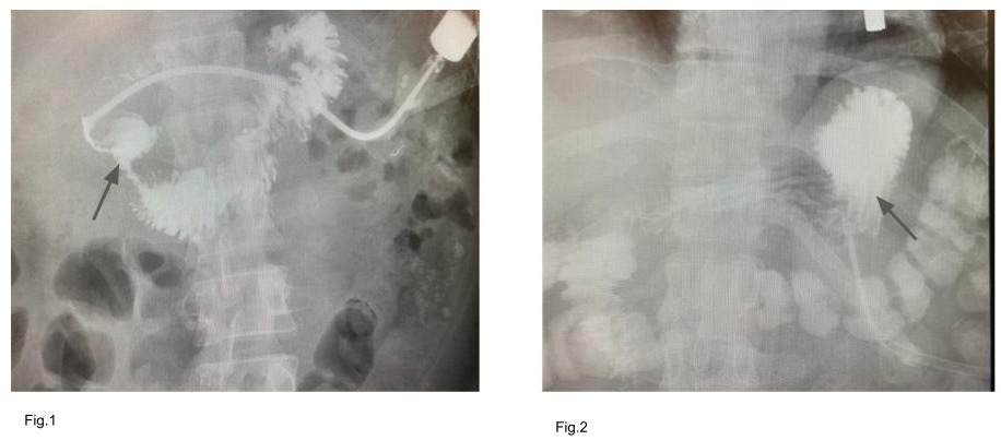
Case Description/Methods: A 43-year-old man with diabetes, traumatic brain injury, stroke, and ulcerative colitis. Underwent PEG tube placement for long-term nutrition and medication use. PEG tube being used uneventfully for more than two years. He presented to hospital with complaints of resistance to tube feeds and abdominal pain for one day. On arrival the patient is in mild abdominal pain, otherwise vital signs were in normal range. On physical examination, he has a 20F PEG tube in the left upper quadrant area. The external bolster is almost close to the feeding port. Unable to pull the PEG tube outwards, it felt like the internal balloon stuck inside the stomach. Even under gentle pressure, the patient was complaining of pain. The imaging of the tube was performed using gastrografin instilled through the tube. Which showed the internal balloon /distal tip of the tube is inside the proximal aspect of the duodenum (Fig 1). It was confirmed that the PEG tube has migrated into the pylorus. At the bedside, deflated the internal balloon and the PEG tube was removed with some resistance. A new 20F PEG tube was inserted through the same track, the internal balloon inflated and the external bolster was secured properly. Post-placement of the new PEG tube, the repeat imaging using gastrografin , showed the internal balloon is within the stomach(Fig 2). Patient was discharged home with proper instructions to prevent and avoid complications of a similar type.
Discussion: The complications associated with PEG tubes range from simple tube clogging to serious buried bumper syndrome. The tube migration is a rare complication, which may occur due to increased peristalsis which could push the internal balloon into the pylorus. Patients with this complication are at risk of gastric outlet obstruction, even block the pancreatic, biliary ducts leading to serious complications like pancreatitis. Our case provides useful insight into the importance of recognizing the tube migration early and treating it appropriately to prevent any further complications and minimize patient suffering.

Figure: Fig.1 Internal balloon is seen inside the proximal aspect of the duodenum, ( arrow); Fig.2 Internal balloon is seen within the stomach. (arrow).
Disclosures:
Mahesh Cheryala indicated no relevant financial relationships.
Yichen Wang indicated no relevant financial relationships.
Sreekant Avula indicated no relevant financial relationships.
Martha Ghosh indicated no relevant financial relationships.
Vamsee Chirumamilla indicated no relevant financial relationships.
Mahesh Cheryala, MD, MRCP1, Yichen Wang, MD2, Sreekant Avula, MD3, Martha Ghosh, MD4, Vamsee Chirumamilla, MD3. B0304 - A Rare Complication of Percutaneous Endoscopic Gastrostomy Tube: A Case Report, ACG 2022 Annual Scientific Meeting Abstracts. Charlotte, NC: American College of Gastroenterology.
