Back


Poster Session B - Monday Morning
Category: Biliary/Pancreas
B0049 - Kaposi Sarcoma: An Unusual Cause of Biliary Obstruction
Monday, October 24, 2022
10:00 AM – 12:00 PM ET
Location: Crown Ballroom

Has Audio

Rebecca Salvo, DO
Valley Hospital Medical Center
Las Vegas, NV
Presenting Author(s)
Rebecca Salvo, DO1, Shahid Wahid, MD2, Vishvinder Sharma, MD1, Scott Diamond, DO1, Alexis Serrano, DO3
1Valley Hospital Medical Center, Las Vegas, NV; 2Kirk Kerkorian School of Medicine at UNLV, Las Vegas, NV; 3Valley Medical Hospital Center, Las Vegas, NV
Introduction: Kaposi sarcoma (KS) is an AIDs defining disease caused by human herpesvirus-8 occurring in setting of severe immunosuppression. AIDS-associated KS affects primarily the skin and the lungs, but biliary tract involvement has been reported. We present an uncommon presentation of disseminated Kaposi sarcoma causing intrabiliary stricturing that resolved after biliary stent placement.
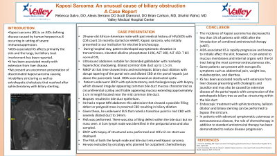
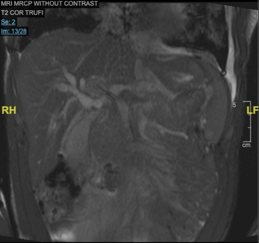
Case Description/Methods: 29-year-old African-American male with past medical history of HIV/AIDS with CD4 count 15 recently started on HAART, Kaposi sarcoma, who initially presented to our institution for elective bronchoscopy. During hospital stay, patient developed elevated alkaline phosphatase 1196, AST 180, ALT 150. He denied any abdominal symptoms. Ultrasound abdomen noted a distended gallbladder with markedly hyperechoic shadowing, dilated common bile duct to 1.5 cm. Of note, patient was previously admitted to a sister hospital with abnormal liver enzymes. MRCP at that time showed intra and extrahepatic biliary duct dilation with abrupt tapering of the portal vein and dilated CBD at the portal hepatis just above the pancreatic head. Patient underwent ERCP with sphincterotomy and cholangioscopy with biopsy, which showed irregular-appearing common bile duct mucosa characterized as circumferential scallop and friable appearing mucosa extending approximately 1 cm in length located near the mid common bile duct. Biopsies resulted in bile duct epithelium. He had a repeat MRI abdomen this admission that showed a possible filling defect or polypoid mass in proximal CBD resulting in biliary dilation Given this, he underwent EUS that noted a transition point in mid CBD with severely dilated duct to 14mm. FNA was performed. There was also a filling defect within the bile duct but no mass seen. A 3cm lymph node was identified in the periportal area and also sampled. ERCP with biopsy of structured area performed and 10Fx12 cm stent was deployed. The FNA of both the lymph node and bile duct returned Kaposi sarcoma. He was evaluated by oncology who planned for outpatient chemotherapy.
Discussion: AIDS-associated KS is known to primarily affect the skin; however, it can extend to internal organs, with the GI tract being the most common extracutaneous site. This may present with non-specific symptoms or as in this case elevated LFTs in a cholestatic pattern. Other times, patients may present with jaundice, cholangitis. Chemotherapy with antiretroviral therapy has been demonstrated to reduce disease progression.

Disclosures:
Rebecca Salvo, DO1, Shahid Wahid, MD2, Vishvinder Sharma, MD1, Scott Diamond, DO1, Alexis Serrano, DO3. B0049 - Kaposi Sarcoma: An Unusual Cause of Biliary Obstruction, ACG 2022 Annual Scientific Meeting Abstracts. Charlotte, NC: American College of Gastroenterology.
1Valley Hospital Medical Center, Las Vegas, NV; 2Kirk Kerkorian School of Medicine at UNLV, Las Vegas, NV; 3Valley Medical Hospital Center, Las Vegas, NV
Introduction: Kaposi sarcoma (KS) is an AIDs defining disease caused by human herpesvirus-8 occurring in setting of severe immunosuppression. AIDS-associated KS affects primarily the skin and the lungs, but biliary tract involvement has been reported. We present an uncommon presentation of disseminated Kaposi sarcoma causing intrabiliary stricturing that resolved after biliary stent placement.
Case Description/Methods: 29-year-old African-American male with past medical history of HIV/AIDS with CD4 count 15 recently started on HAART, Kaposi sarcoma, who initially presented to our institution for elective bronchoscopy. During hospital stay, patient developed elevated alkaline phosphatase 1196, AST 180, ALT 150. He denied any abdominal symptoms. Ultrasound abdomen noted a distended gallbladder with markedly hyperechoic shadowing, dilated common bile duct to 1.5 cm. Of note, patient was previously admitted to a sister hospital with abnormal liver enzymes. MRCP at that time showed intra and extrahepatic biliary duct dilation with abrupt tapering of the portal vein and dilated CBD at the portal hepatis just above the pancreatic head. Patient underwent ERCP with sphincterotomy and cholangioscopy with biopsy, which showed irregular-appearing common bile duct mucosa characterized as circumferential scallop and friable appearing mucosa extending approximately 1 cm in length located near the mid common bile duct. Biopsies resulted in bile duct epithelium. He had a repeat MRI abdomen this admission that showed a possible filling defect or polypoid mass in proximal CBD resulting in biliary dilation Given this, he underwent EUS that noted a transition point in mid CBD with severely dilated duct to 14mm. FNA was performed. There was also a filling defect within the bile duct but no mass seen. A 3cm lymph node was identified in the periportal area and also sampled. ERCP with biopsy of structured area performed and 10Fx12 cm stent was deployed. The FNA of both the lymph node and bile duct returned Kaposi sarcoma. He was evaluated by oncology who planned for outpatient chemotherapy.
Discussion: AIDS-associated KS is known to primarily affect the skin; however, it can extend to internal organs, with the GI tract being the most common extracutaneous site. This may present with non-specific symptoms or as in this case elevated LFTs in a cholestatic pattern. Other times, patients may present with jaundice, cholangitis. Chemotherapy with antiretroviral therapy has been demonstrated to reduce disease progression.

Figure: MRCP with possible filling defect/polypoid mass in proximal CBD resulting in biliary dilation
Disclosures:
Rebecca Salvo indicated no relevant financial relationships.
Shahid Wahid indicated no relevant financial relationships.
Vishvinder Sharma indicated no relevant financial relationships.
Scott Diamond indicated no relevant financial relationships.
Alexis Serrano indicated no relevant financial relationships.
Rebecca Salvo, DO1, Shahid Wahid, MD2, Vishvinder Sharma, MD1, Scott Diamond, DO1, Alexis Serrano, DO3. B0049 - Kaposi Sarcoma: An Unusual Cause of Biliary Obstruction, ACG 2022 Annual Scientific Meeting Abstracts. Charlotte, NC: American College of Gastroenterology.
