Back


Poster Session B - Monday Morning
Category: IBD
B0435 - A Rare Triad of Ulcerative Colitis, Large Vessel Vasculitis and Celiac Disease
Monday, October 24, 2022
10:00 AM – 12:00 PM ET
Location: Crown Ballroom

Has Audio

Ying Zhou, MD
NYU Langone Health
New York, New York
Presenting Author(s)
Mary Guan, MD1, Ying Zhou, MD1, Yamen Homsi, MD2, Shannon Chang, MD1
1NYU Langone Health, New York, NY; 2NYU Langone Health, Brooklyn, NY
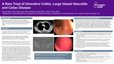
Introduction: Inflammatory bowel disease (IBD) has been associated with large-vessel vasculitis (LVV), with the diagnosis of IBD preceding that of LVV by years. We present for the first time in known literature a triad of concurrent ulcerative colitis (UC), aortitis and celiac disease.
Case Description/Methods: A 58 year old Hispanic man with a history of hypertension and gout presented with two weeks of intractable temporomandibular headaches, and two months of non-bloody diarrhea and weight loss. Physical exam was unremarkable. Labs showed hemoglobin 6.9 g/dL, erythrocyte sedimentation rate 120 mm/hr, C-reactive protein 281 mg/dL and IgA tissue transglutaminase antibody 23 U/mL. ANA, C3, C4, proteinase-3 and myeloperoxidase antibodies were within normal limits.
Colonoscopy showed pancolitis from rectum to ascending colon. The terminal ileum was normal. Abdominal MRI found aortic wall hyperintensity from the renal arteries to common iliac bifurcation. CT angiogram showed wall thickening of the left carotid artery, aortic arch, descending thoracic and abdominal aorta, consistent with vasculitis. Patient was given stress dose steroids with improvement in headache and normalization of ESR and CRP. Temporal artery biopsy was unremarkable. Four months after hospitalization, repeat colonoscopy with duodenal biopsies for celiac disease revealed mild increase in intraepithelial lymphocytes with preserved villous architecture. He was started on a gluten free diet and adalimumab in combination with methotrexate for UC and LVV.
Discussion: About 10 case reports of patients with both UC and either Takayasu (TAK) or giant cell arteritis (GCA) have been described, with UC typically diagnosed 15-45 years before the vasculitis. Vasculitis in the GI tract can mimic IBD, making colonoscopy and biopsy crucial for diagnosis. HLA haplotypes A24, B52, and DR2 are associated with both UC and aortitis and Interleukin-9, observed in temporal arteritis lesions, may be implicated in the pathogenesis of UC. Shared chromosomal variants between patients with UC and celiac disease may explain why IBD risk is up to 9-fold higher in patients with celiac disease. Our patient may have presented with isolated aortitis or an early form of GCA. Methotrexate is used to treat LVV and is combined with an anti-TNF agent to treat UC, as in our case. This is the first known report of co-occurring UC, celiac disease and aortitis; however, whether the three inflammatory conditions are mechanistically related warrants further research.
Disclosures:
Mary Guan, MD1, Ying Zhou, MD1, Yamen Homsi, MD2, Shannon Chang, MD1. B0435 - A Rare Triad of Ulcerative Colitis, Large Vessel Vasculitis and Celiac Disease, ACG 2022 Annual Scientific Meeting Abstracts. Charlotte, NC: American College of Gastroenterology.
1NYU Langone Health, New York, NY; 2NYU Langone Health, Brooklyn, NY
Introduction: Inflammatory bowel disease (IBD) has been associated with large-vessel vasculitis (LVV), with the diagnosis of IBD preceding that of LVV by years. We present for the first time in known literature a triad of concurrent ulcerative colitis (UC), aortitis and celiac disease.
Case Description/Methods: A 58 year old Hispanic man with a history of hypertension and gout presented with two weeks of intractable temporomandibular headaches, and two months of non-bloody diarrhea and weight loss. Physical exam was unremarkable. Labs showed hemoglobin 6.9 g/dL, erythrocyte sedimentation rate 120 mm/hr, C-reactive protein 281 mg/dL and IgA tissue transglutaminase antibody 23 U/mL. ANA, C3, C4, proteinase-3 and myeloperoxidase antibodies were within normal limits.
Colonoscopy showed pancolitis from rectum to ascending colon. The terminal ileum was normal. Abdominal MRI found aortic wall hyperintensity from the renal arteries to common iliac bifurcation. CT angiogram showed wall thickening of the left carotid artery, aortic arch, descending thoracic and abdominal aorta, consistent with vasculitis. Patient was given stress dose steroids with improvement in headache and normalization of ESR and CRP. Temporal artery biopsy was unremarkable. Four months after hospitalization, repeat colonoscopy with duodenal biopsies for celiac disease revealed mild increase in intraepithelial lymphocytes with preserved villous architecture. He was started on a gluten free diet and adalimumab in combination with methotrexate for UC and LVV.
Discussion: About 10 case reports of patients with both UC and either Takayasu (TAK) or giant cell arteritis (GCA) have been described, with UC typically diagnosed 15-45 years before the vasculitis. Vasculitis in the GI tract can mimic IBD, making colonoscopy and biopsy crucial for diagnosis. HLA haplotypes A24, B52, and DR2 are associated with both UC and aortitis and Interleukin-9, observed in temporal arteritis lesions, may be implicated in the pathogenesis of UC. Shared chromosomal variants between patients with UC and celiac disease may explain why IBD risk is up to 9-fold higher in patients with celiac disease. Our patient may have presented with isolated aortitis or an early form of GCA. Methotrexate is used to treat LVV and is combined with an anti-TNF agent to treat UC, as in our case. This is the first known report of co-occurring UC, celiac disease and aortitis; however, whether the three inflammatory conditions are mechanistically related warrants further research.
Disclosures:
Mary Guan indicated no relevant financial relationships.
Ying Zhou indicated no relevant financial relationships.
Yamen Homsi indicated no relevant financial relationships.
Shannon Chang: Abbvie – Consultant. BMS – Consultant. Pfizer – Consultant.
Mary Guan, MD1, Ying Zhou, MD1, Yamen Homsi, MD2, Shannon Chang, MD1. B0435 - A Rare Triad of Ulcerative Colitis, Large Vessel Vasculitis and Celiac Disease, ACG 2022 Annual Scientific Meeting Abstracts. Charlotte, NC: American College of Gastroenterology.
