Back


Poster Session C - Monday Afternoon
Category: IBD
C0430 - Clinical Correlation Recommended: Appendiceal Crohn’s Masquerading as Acute Appendicitis on CT
Monday, October 24, 2022
3:00 PM – 5:00 PM ET
Location: Crown Ballroom

Has Audio

Stacey Rolak, MD, MPH
Mayo Clinic
Rochester, MN
Presenting Author(s)
Stacey Rolak, MD, MPH, Katie A. Dunleavy, MB, BCh, BAO, Philip Hurst, MD, Gary Keeney, MD, Xiao Jing (Iris) Wang, MD
Mayo Clinic, Rochester, MN
Introduction: Granulomatous appendicitis is uncommon, often caused by Crohn’s disease (CD), infiltrative disease (ie sarcoidosis), or infection (ie Yersinia). We report an unusual presentation of CD limited to the appendix.
Case Description/Methods: A 46-year-old male with celiac disease presented to clinic for evaluation of intermittent epigastric and right upper quadrant (RUQ) abdominal pain occurring four times per year. When present, the pain was constant, pressure-like and gradually worsened over a three-day period. He denied nausea or vomiting, but reported non-bloody diarrhea with episodes. His symptoms persisted despite a gluten free diet and normalization of celiac serologies. In the clinic, he was afebrile with normal vital signs. Abdominal exam demonstrated mild tenderness to deep palpation of the RUQ. CT enterography demonstrated inflammation around his appendix, terminal ileum, and right ureter concerning for acute appendicitis (Figure A). Given incongruity of his mild clinical presentation and chronicity of his symptoms, he underwent colonoscopy which showed inflammation of the appendiceal orifice, surrounding cecum, and distal rectum. Biopsies of the rectum and cecum demonstrated active inflammation without chronicity, with a normal terminal ileum. Due to evidence of active inflammation of unknown etiology, he underwent laparoscopic ileocecectomy with anastomosis. Pathology findings were consistent with moderately active Crohn’s colitis isolated to the appendix. He had no signs or symptoms of extra-intestinal manifestations of inflammatory bowel disease. Following surgery, his symptoms resolved and repeat CT six months later demonstrated resolution of inflammation. The patient remains asymptomatic without CD treatment.
Discussion: Isolated appendiceal CD is a rare entity that is most often seen in men in their 20-30s. It typically presents with acute right lower quadrant pain, occasionally with diarrhea, lasting 0-14 days, but can become protracted. Our patient’s history and nontoxic exam did not fit acute appendicitis as initially suspected on CT scan, instead suggesting a chronic process such as CD. There is no consensus regarding surveillance and treatment of isolated appendiceal CD. Recurrence rates after appendectomy are low, suggesting that appendectomy alone is a sufficient treatment for isolated disease. It was therefore recommended that this patient return for further evaluation based on symptoms alone, rather than undergo surveillance.

Disclosures:
Stacey Rolak, MD, MPH, Katie A. Dunleavy, MB, BCh, BAO, Philip Hurst, MD, Gary Keeney, MD, Xiao Jing (Iris) Wang, MD. C0430 - Clinical Correlation Recommended: Appendiceal Crohn’s Masquerading as Acute Appendicitis on CT, ACG 2022 Annual Scientific Meeting Abstracts. Charlotte, NC: American College of Gastroenterology.
Mayo Clinic, Rochester, MN
Introduction: Granulomatous appendicitis is uncommon, often caused by Crohn’s disease (CD), infiltrative disease (ie sarcoidosis), or infection (ie Yersinia). We report an unusual presentation of CD limited to the appendix.
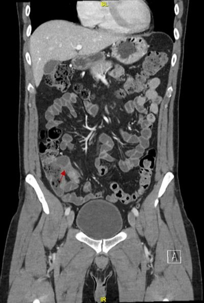
Case Description/Methods: A 46-year-old male with celiac disease presented to clinic for evaluation of intermittent epigastric and right upper quadrant (RUQ) abdominal pain occurring four times per year. When present, the pain was constant, pressure-like and gradually worsened over a three-day period. He denied nausea or vomiting, but reported non-bloody diarrhea with episodes. His symptoms persisted despite a gluten free diet and normalization of celiac serologies. In the clinic, he was afebrile with normal vital signs. Abdominal exam demonstrated mild tenderness to deep palpation of the RUQ. CT enterography demonstrated inflammation around his appendix, terminal ileum, and right ureter concerning for acute appendicitis (Figure A). Given incongruity of his mild clinical presentation and chronicity of his symptoms, he underwent colonoscopy which showed inflammation of the appendiceal orifice, surrounding cecum, and distal rectum. Biopsies of the rectum and cecum demonstrated active inflammation without chronicity, with a normal terminal ileum. Due to evidence of active inflammation of unknown etiology, he underwent laparoscopic ileocecectomy with anastomosis. Pathology findings were consistent with moderately active Crohn’s colitis isolated to the appendix. He had no signs or symptoms of extra-intestinal manifestations of inflammatory bowel disease. Following surgery, his symptoms resolved and repeat CT six months later demonstrated resolution of inflammation. The patient remains asymptomatic without CD treatment.
Discussion: Isolated appendiceal CD is a rare entity that is most often seen in men in their 20-30s. It typically presents with acute right lower quadrant pain, occasionally with diarrhea, lasting 0-14 days, but can become protracted. Our patient’s history and nontoxic exam did not fit acute appendicitis as initially suspected on CT scan, instead suggesting a chronic process such as CD. There is no consensus regarding surveillance and treatment of isolated appendiceal CD. Recurrence rates after appendectomy are low, suggesting that appendectomy alone is a sufficient treatment for isolated disease. It was therefore recommended that this patient return for further evaluation based on symptoms alone, rather than undergo surveillance.

Figure: Figure A. CT Abdomen Pelvis Enterography with IV Contrast demonstrating appendiceal inflammation.
Disclosures:
Stacey Rolak indicated no relevant financial relationships.
Katie Dunleavy indicated no relevant financial relationships.
Philip Hurst indicated no relevant financial relationships.
Gary Keeney indicated no relevant financial relationships.
Xiao Jing (Iris) Wang indicated no relevant financial relationships.
Stacey Rolak, MD, MPH, Katie A. Dunleavy, MB, BCh, BAO, Philip Hurst, MD, Gary Keeney, MD, Xiao Jing (Iris) Wang, MD. C0430 - Clinical Correlation Recommended: Appendiceal Crohn’s Masquerading as Acute Appendicitis on CT, ACG 2022 Annual Scientific Meeting Abstracts. Charlotte, NC: American College of Gastroenterology.

