Back


Poster Session C - Monday Afternoon
Category: Colon
C0125 - Hiding Mantle Cell Lymphoma
Monday, October 24, 2022
3:00 PM – 5:00 PM ET
Location: Crown Ballroom

Has Audio

Tudor Oroian, MD
Walter Reed National Military Medical Center
Bethesda, MD
Presenting Author(s)
Tudor Oroian, MD, Gillian Costa, MD, Allison Bush, MD, Dean Baird, MD, Patrick Young, MD, FACG
Walter Reed National Military Medical Center, Bethesda, MD
Introduction: Primary gastrointestinal lymphomas represent 1-4% of GI malignancies. Mantle cell lymphoma (MCL) is a Non-Hodgkin’s lymphoma that -in the GI tract- is rare, representing fewer than 5% of primary gastrointestinal lymphomas. The clinical course ranges from indolent to aggressive. A patient’s symptoms, imaging, and endoscopic findings can be non-specific, making the diagnosis of MCL challenging. We present a patient with an elusive intestinal lesion that was diagnosed as a primary gastrointestinal MCL.
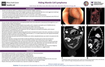
Case Description/Methods: A 74-year-old man with a history of hypothyroidism and prostate cancer treated with prostatectomy presented with polyarthralgia, fatigue, dyspepsia and anorexia with 20-pound weight loss over two months. His physical exam revealed no abnormal findings. Laboratory studies revealed peripheral eosinophilia on complete blood count with an absolute eosinophil count of 1800 cells/mcL. Computed tomography showed a soft tissue lesion in the region of the cecum and ascending colon with multiple right lower quadrant sub-centimeter mesenteric lymph nodes. He underwent subsequent colonoscopy that demonstrated a normal appearing cecum and ascending colon. He was then evaluated with PET, which showed hypermetabolic activity at the ileocecal valve. He underwent a diagnostic laparoscopy which found no evidence of an extraluminal colonic mass nor mass in the mesentery or omentum. Repeat colonoscopy revealed a lesion in the terminal ileum with biopsies noting atypical lymphoid infiltrate with t(11;14)(q13;q32) on FISH analysis and the patient was diagnosed with MCL. Patient’s symptoms resolved and given the asymptomatic and localized nature with isolated gastrointestinal extra-nodal disease he is monitored with serial imaging.
Discussion: Primary gastrointestinal MCL is a rare disease with a variety of clinical presentations. The diagnosis can be challenging as patients who are symptomatic present with vague reports of anorexia, bloating or abdominal pain. Radiographically the lymphoma may or may not be apparent. Endoscopically the MCL can range from normal appearing mucosa to polypoid or ulcerated lesions. In this patient, it is likely that the ileal lesion periodically prolapsed into the colon- explaining the imaging findings. In the initial colonoscopy, the prolapsed segment spontaneously reduced, leaving only the falsely reassuring normal colon. High clinical suspicion based on subsequent imaging led to repeat colonoscopy with ileal intubation and tissue sampling, yielding the diagnosis.
Disclosures:
Tudor Oroian, MD, Gillian Costa, MD, Allison Bush, MD, Dean Baird, MD, Patrick Young, MD, FACG. C0125 - Hiding Mantle Cell Lymphoma, ACG 2022 Annual Scientific Meeting Abstracts. Charlotte, NC: American College of Gastroenterology.
Walter Reed National Military Medical Center, Bethesda, MD
Introduction: Primary gastrointestinal lymphomas represent 1-4% of GI malignancies. Mantle cell lymphoma (MCL) is a Non-Hodgkin’s lymphoma that -in the GI tract- is rare, representing fewer than 5% of primary gastrointestinal lymphomas. The clinical course ranges from indolent to aggressive. A patient’s symptoms, imaging, and endoscopic findings can be non-specific, making the diagnosis of MCL challenging. We present a patient with an elusive intestinal lesion that was diagnosed as a primary gastrointestinal MCL.
Case Description/Methods: A 74-year-old man with a history of hypothyroidism and prostate cancer treated with prostatectomy presented with polyarthralgia, fatigue, dyspepsia and anorexia with 20-pound weight loss over two months. His physical exam revealed no abnormal findings. Laboratory studies revealed peripheral eosinophilia on complete blood count with an absolute eosinophil count of 1800 cells/mcL. Computed tomography showed a soft tissue lesion in the region of the cecum and ascending colon with multiple right lower quadrant sub-centimeter mesenteric lymph nodes. He underwent subsequent colonoscopy that demonstrated a normal appearing cecum and ascending colon. He was then evaluated with PET, which showed hypermetabolic activity at the ileocecal valve. He underwent a diagnostic laparoscopy which found no evidence of an extraluminal colonic mass nor mass in the mesentery or omentum. Repeat colonoscopy revealed a lesion in the terminal ileum with biopsies noting atypical lymphoid infiltrate with t(11;14)(q13;q32) on FISH analysis and the patient was diagnosed with MCL. Patient’s symptoms resolved and given the asymptomatic and localized nature with isolated gastrointestinal extra-nodal disease he is monitored with serial imaging.
Discussion: Primary gastrointestinal MCL is a rare disease with a variety of clinical presentations. The diagnosis can be challenging as patients who are symptomatic present with vague reports of anorexia, bloating or abdominal pain. Radiographically the lymphoma may or may not be apparent. Endoscopically the MCL can range from normal appearing mucosa to polypoid or ulcerated lesions. In this patient, it is likely that the ileal lesion periodically prolapsed into the colon- explaining the imaging findings. In the initial colonoscopy, the prolapsed segment spontaneously reduced, leaving only the falsely reassuring normal colon. High clinical suspicion based on subsequent imaging led to repeat colonoscopy with ileal intubation and tissue sampling, yielding the diagnosis.
Disclosures:
Tudor Oroian indicated no relevant financial relationships.
Gillian Costa indicated no relevant financial relationships.
Allison Bush indicated no relevant financial relationships.
Dean Baird indicated no relevant financial relationships.
Patrick Young indicated no relevant financial relationships.
Tudor Oroian, MD, Gillian Costa, MD, Allison Bush, MD, Dean Baird, MD, Patrick Young, MD, FACG. C0125 - Hiding Mantle Cell Lymphoma, ACG 2022 Annual Scientific Meeting Abstracts. Charlotte, NC: American College of Gastroenterology.
