Back


Poster Session C - Monday Afternoon
Category: Interventional Endoscopy
C0483 - Novel Use of Lumen Apposing Metal Stent for Management of Gastrointestinal Perforations
Monday, October 24, 2022
3:00 PM – 5:00 PM ET
Location: Crown Ballroom

Has Audio

Fouzia Oza, MD
Wright Center for Graduate Medical Education
Scranton, Pennsylvania
Presenting Author(s)
Award: Presidential Poster Award
Fouzia Oza, MD1, Hemant Goyal, MD2, Jelena Surla, MD3, Vikas Khurana, MD3, Aman Ali, MD3
1Wright Center for Graduate Medical Education, Scranton, PA; 2University of Texas McGovern Medical School, Houston, TX; 3The Wright Center for Graduate Medical Education, Scranton, PA
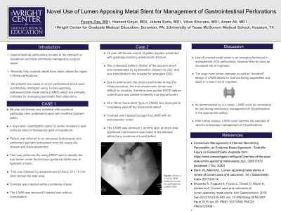
Introduction: Gastrointestinal perforations located in the stomach or duodenum are most commonly managed by surgical repair. Recently, some cases have been reported in which fully covered stents have been utilized for repair in these perforations. To our knowledge, lumen-apposing self-expandable metal stents (LAMS) have not been used before for primary or secondary management of gastroduodenal perforations. Herein, we present 2 cases of perforations which were successfully managed using LAMS which are primarily indicated for managing pancreatic fluid collections.
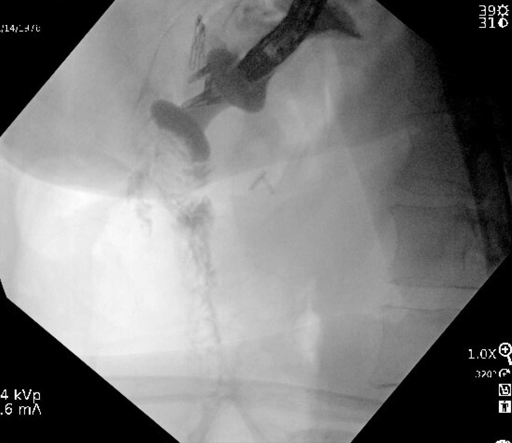
Case Description/Methods: Case 1: A 45-year-old female with history of gastric bypass underwent outpatient balloon dilation for gastrojejunostomy anastomotic stricture which was complicated by a perforation. Patient was transferred to the hospital and underwent emergent EGD. True anastomotic lumen was difficult to visualize due to edema and clip closure placed during the initial procedure. Wire Guided ERCP balloons under fluoro were able to identify true jejunal lumen. A 20 x 10mm Axios stent (type of LAMS) was deployed to completely seal off the transmural defect. Contrast was injected through the LAMS with no extravasation noted. The LAMS was removed 2 months later at which time significant improvement was noted in the stricture without any evidence of mural defect.
Case 2:
A 66-year-old female was admitted with duodenal perforation who underwent repair with modified Graham patch. 4 days later, Gastrograffin upper GI series revealed leak at the junction of first/second part of duodenum. Patient was referred to advanced endoscopist who performed egd with endoscopic over the scope clip closure and Axios placement . This was performed by using ERCP wire to identifiy the true lumen under fluoroscopic guidance all the way to ligament of trietz. This was followed by advancement of Axios 20 x 10 mm stent across the leak area. Contrast was injected without evidence of leak. The LAMS was removed 6 weeks later without complication.
Discussion: Use of covered metal stents is an emerging technique in management of GI perforations, however they do have an increased risk of migration. The “dumbbell” design of LAMS allows for dual anchoring capabilities and leads to a lower risk of migration. As demonstrated by our cases, LAMS could be considered for use during endoscopic management of GI perforations in the appropriate setting. With further studies, LAMS could become the standard of care for endoscopic management of GI perforations.

Disclosures:
Fouzia Oza, MD1, Hemant Goyal, MD2, Jelena Surla, MD3, Vikas Khurana, MD3, Aman Ali, MD3. C0483 - Novel Use of Lumen Apposing Metal Stent for Management of Gastrointestinal Perforations, ACG 2022 Annual Scientific Meeting Abstracts. Charlotte, NC: American College of Gastroenterology.
Fouzia Oza, MD1, Hemant Goyal, MD2, Jelena Surla, MD3, Vikas Khurana, MD3, Aman Ali, MD3
1Wright Center for Graduate Medical Education, Scranton, PA; 2University of Texas McGovern Medical School, Houston, TX; 3The Wright Center for Graduate Medical Education, Scranton, PA
Introduction: Gastrointestinal perforations located in the stomach or duodenum are most commonly managed by surgical repair. Recently, some cases have been reported in which fully covered stents have been utilized for repair in these perforations. To our knowledge, lumen-apposing self-expandable metal stents (LAMS) have not been used before for primary or secondary management of gastroduodenal perforations. Herein, we present 2 cases of perforations which were successfully managed using LAMS which are primarily indicated for managing pancreatic fluid collections.
Case Description/Methods: Case 1: A 45-year-old female with history of gastric bypass underwent outpatient balloon dilation for gastrojejunostomy anastomotic stricture which was complicated by a perforation. Patient was transferred to the hospital and underwent emergent EGD. True anastomotic lumen was difficult to visualize due to edema and clip closure placed during the initial procedure. Wire Guided ERCP balloons under fluoro were able to identify true jejunal lumen. A 20 x 10mm Axios stent (type of LAMS) was deployed to completely seal off the transmural defect. Contrast was injected through the LAMS with no extravasation noted. The LAMS was removed 2 months later at which time significant improvement was noted in the stricture without any evidence of mural defect.
Case 2:
A 66-year-old female was admitted with duodenal perforation who underwent repair with modified Graham patch. 4 days later, Gastrograffin upper GI series revealed leak at the junction of first/second part of duodenum. Patient was referred to advanced endoscopist who performed egd with endoscopic over the scope clip closure and Axios placement . This was performed by using ERCP wire to identifiy the true lumen under fluoroscopic guidance all the way to ligament of trietz. This was followed by advancement of Axios 20 x 10 mm stent across the leak area. Contrast was injected without evidence of leak. The LAMS was removed 6 weeks later without complication.
Discussion: Use of covered metal stents is an emerging technique in management of GI perforations, however they do have an increased risk of migration. The “dumbbell” design of LAMS allows for dual anchoring capabilities and leads to a lower risk of migration. As demonstrated by our cases, LAMS could be considered for use during endoscopic management of GI perforations in the appropriate setting. With further studies, LAMS could become the standard of care for endoscopic management of GI perforations.

Figure: 20 mm x 10 mm LAMS deployed across the gastrojejunal anastomosis
Disclosures:
Fouzia Oza indicated no relevant financial relationships.
Hemant Goyal: Aimloxy LLC – Consultant.
Jelena Surla indicated no relevant financial relationships.
Vikas Khurana: Aimloxy LLC – Owner/Ownership Interest.
Aman Ali indicated no relevant financial relationships.
Fouzia Oza, MD1, Hemant Goyal, MD2, Jelena Surla, MD3, Vikas Khurana, MD3, Aman Ali, MD3. C0483 - Novel Use of Lumen Apposing Metal Stent for Management of Gastrointestinal Perforations, ACG 2022 Annual Scientific Meeting Abstracts. Charlotte, NC: American College of Gastroenterology.


