Back


Poster Session A - Sunday Afternoon
Category: Liver
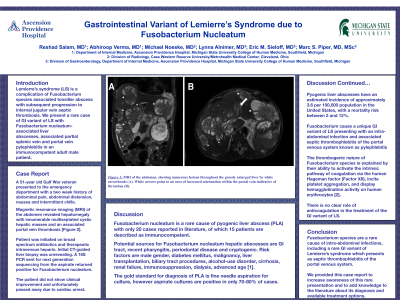
A0577 - Gastrointestinal Variant of Lemierre’s Syndrome Due to Fusobacterium Nucleatum: A Case Report
Sunday, October 23, 2022
5:00 PM – 7:00 PM ET
Location: Crown Ballroom

Has Audio
- RS
Reshad Salam, MD
Ascension Providence Hospital
Southfield, Michigan
Presenting Author(s)
Reshad Salam, MD, Abhiroop Verma, MD, Michael Noeske, MD, Lynna Alnimer, MD, Eric M. Sieloff, MD, Marc S. Piper, MD
Ascension Providence Hospital, Southfield, MI
Introduction: Fusobacterium species are an extremely uncommon cause of pyogenic liver abscess (PLA) and are rarely isolated in the clinical setting. Herein, we report a rare case of cryptogenic Fusobacterium nucleatum-associated liver abscess and septic thrombophlebitis in an apparently immunocompetent patient.
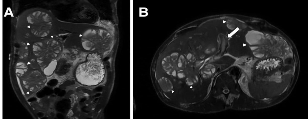
Case Description/Methods: A 51-year-old male patient presented to the emergency department with a two-week history of abdominal pain, distension, nausea and chills. His physical exam revealed a distended abdomen with tenderness to palpation over the right upper and lower quadrants, and palpable hepatomegaly. Laboratory evaluation revealed WBC of 13,360 cells/mm3, lactic acid of 5.2, hemoglobin 8.0 g/dL, ALT of 73 IU/L, AST of 171 IU/L, ALP 625 IU/L, total bilirubin of 1.9 mg/dL and direct bilirubin 1.3 mg/dL. Magnetic resonance imaging (MRI) of the abdomen with and without contrast revealed innumerable multiseptated cystic hepatic masses with an associated portal vein thrombosis. The largest of these cystic lesions was measured at 9.0 cm x 5.4 cm. Patient received empiric intravenous antibiotics and therapeutic intravenous heparin. He underwent a CT-guided liver biopsy with aspiration of abscess material. Blood cultures and aspirate culture were negative. Next generation sequencing 16S PCR of the aspirate was positive for Fusobacterium nucleatum. Unfortunately, the patient passed away due to cardiac arrest before the etiology of the liver lesions could be established.
Discussion: Fusobacterium nucleatum is a rare cause of PLAs with only 20 cases reported in literature. Risk factors for development include recent pharyngitis, periodontal disease or otherwise cryptogenic. Fusobacterium can cause a unique GI variant of Lemierre’s syndrome (LS) presenting with an intra-abdominal infection and associated septic thrombophlebitis of the portal venous system known as pylephlebitis.
Main presenting symptoms in most patients include fever, chills, right upper quadrant abdominal pain, vomiting and shortness of breath. The gold standard for diagnosis of PLA is fine needle aspiration for culture, however aspirate cultures are positive in only 70-80% of cases. Ribosomal RNA (rRNA) gene PCR can be used for detection and identification of bacterial pathogens, as shown in this case. We provided this case report to increase awareness of Fusobacterium- species associated GI variant of LS and to add knowledge to the literature about its presentation, diagnosis, and available treatment options.

Disclosures:
Reshad Salam, MD, Abhiroop Verma, MD, Michael Noeske, MD, Lynna Alnimer, MD, Eric M. Sieloff, MD, Marc S. Piper, MD. A0577 - Gastrointestinal Variant of Lemierre’s Syndrome Due to Fusobacterium Nucleatum: A Case Report, ACG 2022 Annual Scientific Meeting Abstracts. Charlotte, NC: American College of Gastroenterology.
Ascension Providence Hospital, Southfield, MI
Introduction: Fusobacterium species are an extremely uncommon cause of pyogenic liver abscess (PLA) and are rarely isolated in the clinical setting. Herein, we report a rare case of cryptogenic Fusobacterium nucleatum-associated liver abscess and septic thrombophlebitis in an apparently immunocompetent patient.
Case Description/Methods: A 51-year-old male patient presented to the emergency department with a two-week history of abdominal pain, distension, nausea and chills. His physical exam revealed a distended abdomen with tenderness to palpation over the right upper and lower quadrants, and palpable hepatomegaly. Laboratory evaluation revealed WBC of 13,360 cells/mm3, lactic acid of 5.2, hemoglobin 8.0 g/dL, ALT of 73 IU/L, AST of 171 IU/L, ALP 625 IU/L, total bilirubin of 1.9 mg/dL and direct bilirubin 1.3 mg/dL. Magnetic resonance imaging (MRI) of the abdomen with and without contrast revealed innumerable multiseptated cystic hepatic masses with an associated portal vein thrombosis. The largest of these cystic lesions was measured at 9.0 cm x 5.4 cm. Patient received empiric intravenous antibiotics and therapeutic intravenous heparin. He underwent a CT-guided liver biopsy with aspiration of abscess material. Blood cultures and aspirate culture were negative. Next generation sequencing 16S PCR of the aspirate was positive for Fusobacterium nucleatum. Unfortunately, the patient passed away due to cardiac arrest before the etiology of the liver lesions could be established.
Discussion: Fusobacterium nucleatum is a rare cause of PLAs with only 20 cases reported in literature. Risk factors for development include recent pharyngitis, periodontal disease or otherwise cryptogenic. Fusobacterium can cause a unique GI variant of Lemierre’s syndrome (LS) presenting with an intra-abdominal infection and associated septic thrombophlebitis of the portal venous system known as pylephlebitis.
Main presenting symptoms in most patients include fever, chills, right upper quadrant abdominal pain, vomiting and shortness of breath. The gold standard for diagnosis of PLA is fine needle aspiration for culture, however aspirate cultures are positive in only 70-80% of cases. Ribosomal RNA (rRNA) gene PCR can be used for detection and identification of bacterial pathogens, as shown in this case. We provided this case report to increase awareness of Fusobacterium- species associated GI variant of LS and to add knowledge to the literature about its presentation, diagnosis, and available treatment options.

Figure: MRI of the abdomen, showing numerous cystic hepatic lesions throughout the grossly enlarged liver indicated by white arrowheads (A). White arrows point to an area of increased attenuation within the portal vein indicative of thrombus (B).
Disclosures:
Reshad Salam indicated no relevant financial relationships.
Abhiroop Verma indicated no relevant financial relationships.
Michael Noeske indicated no relevant financial relationships.
Lynna Alnimer indicated no relevant financial relationships.
Eric Sieloff indicated no relevant financial relationships.
Marc Piper indicated no relevant financial relationships.
Reshad Salam, MD, Abhiroop Verma, MD, Michael Noeske, MD, Lynna Alnimer, MD, Eric M. Sieloff, MD, Marc S. Piper, MD. A0577 - Gastrointestinal Variant of Lemierre’s Syndrome Due to Fusobacterium Nucleatum: A Case Report, ACG 2022 Annual Scientific Meeting Abstracts. Charlotte, NC: American College of Gastroenterology.
