Back


Poster Session C - Monday Afternoon
Category: Esophagus
C0227 - Dysphagia Lusoria Secondary to an Aberrant Left Subclavian Artery
Monday, October 24, 2022
3:00 PM – 5:00 PM ET
Location: Crown Ballroom

Has Audio

Aizaz Khan, MD
University of Massachusetts Medical School - Baystate
Springfield, MA
Presenting Author(s)
Kevin Groudan, MD1, Aizaz Khan, MD1, Logan Striplin, MD2, Sherin Samuel, DO3, Syed Hamza Sohail, MD1
1University of Massachusetts Medical School - Baystate, Springfield, MA; 2University of Massachusetts Chan Medical School - Baystate, Springfield, MA; 3UMass Chan Medical School - Baystate, Springfield, MA
Introduction: Dysphagia lusoria, also known as Bayford-Autenrieth dysphagia, is a rare condition with prevalence of approximately 0.5%. First reported by David Bayford in 1790, it is defined as dysphagia secondary to extrinsic compression of the esophagus as a result of congenital abnormality of the aortic arch and its branches. We report a case of Dysphagia lusoria in a patient with congenital heart syndrome.
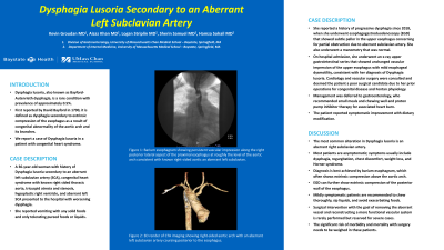
Case Description/Methods: A 36-year-old woman with history of Dysphagia lusoria secondary to an aberrant left subclavian artery (SCA), congenital heart syndrome with known right sided thoracic aorta, tricuspid atresia and stenosis, hypoplastic right ventricle, and aberrant left SCA presented to the hospital with worsening dysphagia. She reported vomiting with any solid foods and only tolerating pureed foods or liquids. She reported a history of progressive dysphagia since 2018, when she underwent an esophagogastroduodenoscopy (EGD) that showed subtle pallor in the upper esophagus concerning for partial obstruction due to aberrant subclavian artery. She also underwent a manometry that was normal.
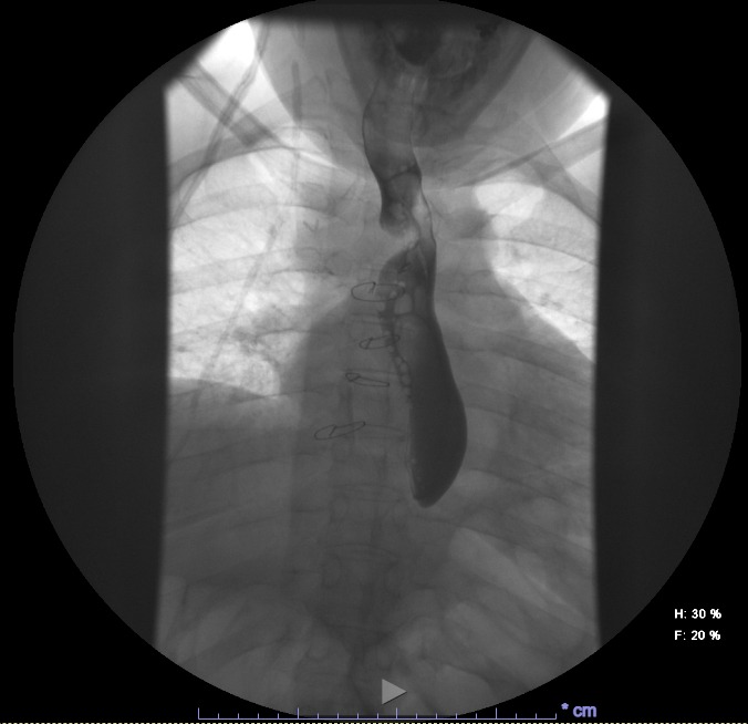
On hospital admission, she underwent an x-ray upper gastrointestinal series that showed unchanged vascular impression of the upper esophagus with mild esophageal dysmotility, consistent with her diagnosis of Dysphagia lusoria. Cardiology and vascular surgery were consulted and deemed the patient a poor surgical candidate due to her prior operations for congenital disease and fontan physiology. Management was deferred to gastroenterology, who recommended small meals and chewing well and proton pump inhibitor therapy for associated heart burn. The patient reported symptomatic improvement with dietary modification.
Discussion: The most common alteration in Dysphagia lusoria is an aberrant right subclavian artery. Most patients are asymptomatic; symptoms usually include dysphagia, regurgitation, chest discomfort, weight loss, and Horner syndrome. Diagnosis is best achieved by barium esophagram, which often shows extrinsic compression above the aortic arch. EGD can further show extrinsic compression of the posterior wall of the esophagus. Mildly symptomatic patients are recommended to chew thoroughly, sip liquids, and avoid exacerbating foods. Surgical intervention with the goal of removing the aberrant vessel and reconstructing a more functional vascular system is rarely performed but reserved for severe cases. The significant risk of morbidity and mortality with surgery needs to be weighed in these patients.

Disclosures:
Kevin Groudan, MD1, Aizaz Khan, MD1, Logan Striplin, MD2, Sherin Samuel, DO3, Syed Hamza Sohail, MD1. C0227 - Dysphagia Lusoria Secondary to an Aberrant Left Subclavian Artery, ACG 2022 Annual Scientific Meeting Abstracts. Charlotte, NC: American College of Gastroenterology.
1University of Massachusetts Medical School - Baystate, Springfield, MA; 2University of Massachusetts Chan Medical School - Baystate, Springfield, MA; 3UMass Chan Medical School - Baystate, Springfield, MA
Introduction: Dysphagia lusoria, also known as Bayford-Autenrieth dysphagia, is a rare condition with prevalence of approximately 0.5%. First reported by David Bayford in 1790, it is defined as dysphagia secondary to extrinsic compression of the esophagus as a result of congenital abnormality of the aortic arch and its branches. We report a case of Dysphagia lusoria in a patient with congenital heart syndrome.
Case Description/Methods: A 36-year-old woman with history of Dysphagia lusoria secondary to an aberrant left subclavian artery (SCA), congenital heart syndrome with known right sided thoracic aorta, tricuspid atresia and stenosis, hypoplastic right ventricle, and aberrant left SCA presented to the hospital with worsening dysphagia. She reported vomiting with any solid foods and only tolerating pureed foods or liquids. She reported a history of progressive dysphagia since 2018, when she underwent an esophagogastroduodenoscopy (EGD) that showed subtle pallor in the upper esophagus concerning for partial obstruction due to aberrant subclavian artery. She also underwent a manometry that was normal.
On hospital admission, she underwent an x-ray upper gastrointestinal series that showed unchanged vascular impression of the upper esophagus with mild esophageal dysmotility, consistent with her diagnosis of Dysphagia lusoria. Cardiology and vascular surgery were consulted and deemed the patient a poor surgical candidate due to her prior operations for congenital disease and fontan physiology. Management was deferred to gastroenterology, who recommended small meals and chewing well and proton pump inhibitor therapy for associated heart burn. The patient reported symptomatic improvement with dietary modification.
Discussion: The most common alteration in Dysphagia lusoria is an aberrant right subclavian artery. Most patients are asymptomatic; symptoms usually include dysphagia, regurgitation, chest discomfort, weight loss, and Horner syndrome. Diagnosis is best achieved by barium esophagram, which often shows extrinsic compression above the aortic arch. EGD can further show extrinsic compression of the posterior wall of the esophagus. Mildly symptomatic patients are recommended to chew thoroughly, sip liquids, and avoid exacerbating foods. Surgical intervention with the goal of removing the aberrant vessel and reconstructing a more functional vascular system is rarely performed but reserved for severe cases. The significant risk of morbidity and mortality with surgery needs to be weighed in these patients.

Figure: X-ray upper gastrointestinal series showing vascular impression of the upper esophagus secondary to aberrant left subclavian artery
Disclosures:
Kevin Groudan indicated no relevant financial relationships.
Aizaz Khan indicated no relevant financial relationships.
Logan Striplin indicated no relevant financial relationships.
Sherin Samuel indicated no relevant financial relationships.
Syed Hamza Sohail indicated no relevant financial relationships.
Kevin Groudan, MD1, Aizaz Khan, MD1, Logan Striplin, MD2, Sherin Samuel, DO3, Syed Hamza Sohail, MD1. C0227 - Dysphagia Lusoria Secondary to an Aberrant Left Subclavian Artery, ACG 2022 Annual Scientific Meeting Abstracts. Charlotte, NC: American College of Gastroenterology.
