Back


Poster Session B - Monday Morning
Category: Colon
B0159 - A Case of Anorectal Junction Melanoma
Monday, October 24, 2022
10:00 AM – 12:00 PM ET
Location: Crown Ballroom

Has Audio

Anya Ramrakhiani
Canterbury High School
Roanoke, IN
Presenting Author(s)
Anya Ramrakhiani, 1, Ashish Sharma, MD2
1Canterbury High School, Roanoke, IN; 2Lutheran Medical Group, Fort Wayne, IN
Introduction: Malignant melanoma of the rectum is an extremely rare disease that is aggressive in nature and often at an advanced stage at the time of diagnosis. We present a case of malignant melanoma of the rectum that was diagnosed on endoscopic polypectomy and staged with endoscopic ultrasound.
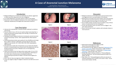
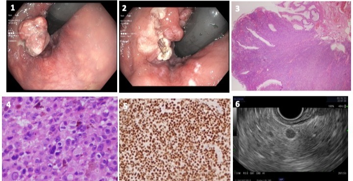
Case Description/Methods: A 66 year-old, asymptomatic, Caucasian female underwent a surveillance colonoscopy. She was noted to have a 20 mm semi-pedunculated rectal polyp (fig.1), which was piecemeal resected using a hot snare (fig. 2). Resection and retrieval were complete. Pathology showed malignant melanoma involving the anal and anorectal junction mucosa (fig. 3 and 4). The tumor approached the inked resection margin. Immunohistochemical stains were positive for Sox-10 (diffuse and strong) (fig. 5), Melan-A (focal), S-100 (focal), and tyrosinase (focal). Stains were negative for pan keratin, CK7, CK20, CDX2, P40, CD56, synaptophysin, CD45, and CEA. EUS performed 3 weeks later showed linear scar just above the dentate line with no residual polyp tissue. Rectal wall at the polypectomy site was thickened without any tumor infiltration and a normal appearing muscular propria. Patient was noted to have two hypoechoic lymph nodes 11.4 mm X 8.5 mm and 6.4 mm X 6.0 mm, 8 cm from anal verge (fig. 6). Fine needle Aspiration biopsy of the lymph nodes was performed. Pathology from the lymph node showed presence of malignant melanoma. Immunostain positive for MART-1 and SOX-10 but negative for HMB-45. PET/CT did not show any regional or distant metastatic disease. Tumor was staged as Stage III malignant melanoma.
Discussion: Malignant melanoma of the distal rectum and anorectal junction is an aggressive and a rare presentation of this disease. It is mostly diagnosed in Caucasian women in fifth to sixth decade. Melanocytes are located at the anal transition zone and squamous zone with most anorectal melanomas arising from the dentate line and located at the anal verge or in the anal canal. Due to the hidden location and late onset of symptoms, many of these tumors are advanced by the time of diagnosis. As this case illustrates, tumors are often 20 mm or bigger in size with nodal involvement. Hence the five year disease free survival in patients with metastatic disease is only 16%.

Disclosures:
Anya Ramrakhiani, 1, Ashish Sharma, MD2. B0159 - A Case of Anorectal Junction Melanoma, ACG 2022 Annual Scientific Meeting Abstracts. Charlotte, NC: American College of Gastroenterology.
1Canterbury High School, Roanoke, IN; 2Lutheran Medical Group, Fort Wayne, IN
Introduction: Malignant melanoma of the rectum is an extremely rare disease that is aggressive in nature and often at an advanced stage at the time of diagnosis. We present a case of malignant melanoma of the rectum that was diagnosed on endoscopic polypectomy and staged with endoscopic ultrasound.
Case Description/Methods: A 66 year-old, asymptomatic, Caucasian female underwent a surveillance colonoscopy. She was noted to have a 20 mm semi-pedunculated rectal polyp (fig.1), which was piecemeal resected using a hot snare (fig. 2). Resection and retrieval were complete. Pathology showed malignant melanoma involving the anal and anorectal junction mucosa (fig. 3 and 4). The tumor approached the inked resection margin. Immunohistochemical stains were positive for Sox-10 (diffuse and strong) (fig. 5), Melan-A (focal), S-100 (focal), and tyrosinase (focal). Stains were negative for pan keratin, CK7, CK20, CDX2, P40, CD56, synaptophysin, CD45, and CEA. EUS performed 3 weeks later showed linear scar just above the dentate line with no residual polyp tissue. Rectal wall at the polypectomy site was thickened without any tumor infiltration and a normal appearing muscular propria. Patient was noted to have two hypoechoic lymph nodes 11.4 mm X 8.5 mm and 6.4 mm X 6.0 mm, 8 cm from anal verge (fig. 6). Fine needle Aspiration biopsy of the lymph nodes was performed. Pathology from the lymph node showed presence of malignant melanoma. Immunostain positive for MART-1 and SOX-10 but negative for HMB-45. PET/CT did not show any regional or distant metastatic disease. Tumor was staged as Stage III malignant melanoma.
Discussion: Malignant melanoma of the distal rectum and anorectal junction is an aggressive and a rare presentation of this disease. It is mostly diagnosed in Caucasian women in fifth to sixth decade. Melanocytes are located at the anal transition zone and squamous zone with most anorectal melanomas arising from the dentate line and located at the anal verge or in the anal canal. Due to the hidden location and late onset of symptoms, many of these tumors are advanced by the time of diagnosis. As this case illustrates, tumors are often 20 mm or bigger in size with nodal involvement. Hence the five year disease free survival in patients with metastatic disease is only 16%.

Figure: Fig. 1: Colonoscopy image showing rectal polyp
Fig. 2: Colonoscopy image showing post rectal polypectomy site
Fig. 3: Pathology showing malignant melanoma involving the anal and anorectal junction mucosa
Fig. 4: Pathology showing sheets of melanocytic tumor cells
Fig. 5: Pathology showing brown nuclear staining with SOX-10 immunoperoxidase marker
Fig. 6: EUS image showing hypoechoic lymph node
Disclosures:
Anya Ramrakhiani indicated no relevant financial relationships.
Ashish Sharma indicated no relevant financial relationships.
Anya Ramrakhiani, 1, Ashish Sharma, MD2. B0159 - A Case of Anorectal Junction Melanoma, ACG 2022 Annual Scientific Meeting Abstracts. Charlotte, NC: American College of Gastroenterology.
