Back


Poster Session B - Monday Morning
Category: Esophagus
B0234 - Epidermoid Metaplasia: An Endoscopic Surveillance Dilemma
Monday, October 24, 2022
10:00 AM – 12:00 PM ET
Location: Crown Ballroom

Has Audio

Robert E. Spiller, DO, MS
Madigan Army Medical Center
Tacoma, WA
Presenting Author(s)
Robert E. Spiller, DO, MS, Jonathan Francis, MD
Madigan Army Medical Center, Tacoma, WA
Introduction: Oral leukoplakia often presents as white patches on oral mucosa and carries a prevalence of 1.5-4.3% within the general population. It is a potentially malignant disorder with its own set of management principles regarding surveillance vs excision/ablation. An analogous entity within the esophagus, epidermoid metaplasia is a rare diagnosis (0.2% of 1,048 consecutive esophageal biopsies) in which the squamous epithelium of the esophagus is replaced with a layer that resembles the epidermis of the skin. It has similar risk factors to oral leukoplakia and presents in middle-aged to elderly patients with a history of smoking and/or alcohol intake. Similar to oral leukoplakia, it has been characterized as a potentially malignant disorder and has been associated with adjacent areas of high grade dysplasia and/or esophageal squamous cell carcinoma.
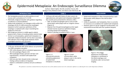
Case Description/Methods: A 69-year-old female with active tobacco use presented for GERD and globus sensation. Esophagogastroduodenoscopy (EGD) showed white plaques with an undulated appearance to the mid-to-distal esophagus (Figures 1a/1b). Mid-esophageal biopsy specimens noted a granular cell layer and hyperkeratosis, consistent with epidermoid metaplasia. EGD one year later showed similar endoscopic findings without evidence of dysplasia. Patient is scheduled to continue annual surveillance EGD.
A 43-year-old-male with history of colon cancer s/p sigmoidectomy presented for refractory reflux. EGD showed Los Angeles Grade B esophagitis with an area of well-demarcated, white plaque with an irregular contour (Figure 1c). Biopsies from the gastroesophageal junction showed reactive squamous epithelial changes, including epidermoid metaplasia. His omeprazole was up-titrated to twice daily with improvement of symptoms. Patient is pending surveillance EGD in 1 year.
Discussion: Epidermoid metaplasia is often characterized by a well-demarcated, white plaque in the mid-to-distal esophagus. Unlike its equivalent, oral leukoplakia, there is currently no consensus on surveillance for epidermoid metaplasia. However, given its risk for malignant transformation, we recommend annual EGD with four quadrant biopsies.

Disclosures:
Robert E. Spiller, DO, MS, Jonathan Francis, MD. B0234 - Epidermoid Metaplasia: An Endoscopic Surveillance Dilemma, ACG 2022 Annual Scientific Meeting Abstracts. Charlotte, NC: American College of Gastroenterology.
Madigan Army Medical Center, Tacoma, WA
Introduction: Oral leukoplakia often presents as white patches on oral mucosa and carries a prevalence of 1.5-4.3% within the general population. It is a potentially malignant disorder with its own set of management principles regarding surveillance vs excision/ablation. An analogous entity within the esophagus, epidermoid metaplasia is a rare diagnosis (0.2% of 1,048 consecutive esophageal biopsies) in which the squamous epithelium of the esophagus is replaced with a layer that resembles the epidermis of the skin. It has similar risk factors to oral leukoplakia and presents in middle-aged to elderly patients with a history of smoking and/or alcohol intake. Similar to oral leukoplakia, it has been characterized as a potentially malignant disorder and has been associated with adjacent areas of high grade dysplasia and/or esophageal squamous cell carcinoma.
Case Description/Methods: A 69-year-old female with active tobacco use presented for GERD and globus sensation. Esophagogastroduodenoscopy (EGD) showed white plaques with an undulated appearance to the mid-to-distal esophagus (Figures 1a/1b). Mid-esophageal biopsy specimens noted a granular cell layer and hyperkeratosis, consistent with epidermoid metaplasia. EGD one year later showed similar endoscopic findings without evidence of dysplasia. Patient is scheduled to continue annual surveillance EGD.
A 43-year-old-male with history of colon cancer s/p sigmoidectomy presented for refractory reflux. EGD showed Los Angeles Grade B esophagitis with an area of well-demarcated, white plaque with an irregular contour (Figure 1c). Biopsies from the gastroesophageal junction showed reactive squamous epithelial changes, including epidermoid metaplasia. His omeprazole was up-titrated to twice daily with improvement of symptoms. Patient is pending surveillance EGD in 1 year.
Discussion: Epidermoid metaplasia is often characterized by a well-demarcated, white plaque in the mid-to-distal esophagus. Unlike its equivalent, oral leukoplakia, there is currently no consensus on surveillance for epidermoid metaplasia. However, given its risk for malignant transformation, we recommend annual EGD with four quadrant biopsies.

Figure: Figure 1a. An undulating, shaggy appearance can be seen, characteristic of epidermoid metaplasia.
Figure 1b. A clearly demarcated plaque is visualized in the mid to distal esophagus.
Figure 1c. Narrow band imaging details the white plaque in the distal esophagus.
Figure 1b. A clearly demarcated plaque is visualized in the mid to distal esophagus.
Figure 1c. Narrow band imaging details the white plaque in the distal esophagus.
Disclosures:
Robert Spiller indicated no relevant financial relationships.
Jonathan Francis indicated no relevant financial relationships.
Robert E. Spiller, DO, MS, Jonathan Francis, MD. B0234 - Epidermoid Metaplasia: An Endoscopic Surveillance Dilemma, ACG 2022 Annual Scientific Meeting Abstracts. Charlotte, NC: American College of Gastroenterology.
