Back


Poster Session C - Monday Afternoon
Category: Liver
C0569 - Hepatocellular Carcinoma Presenting as a Clival Mass
Monday, October 24, 2022
3:00 PM – 5:00 PM ET
Location: Crown Ballroom

Has Audio

Hunza Chaudhry, MD
UCSF-Fresno
Fresno, CA
Presenting Author(s)
Hunza Chaudhry, MD1, Sundeep Bekal, MD2, Alakh Gulati, MD2, Hongqi Peng, MD3, Marina Roytman, MD2
1UCSF-Fresno, Fresno, CA; 2UCSF Fresno, Fresno, CA; 3Community Regional Medical Center, Fresno, CA
Introduction: Hepatocellular carcinoma (HCC) comprises the majority of primary liver cancer and has a poor prognosis. Clivus metastasis is rare with only a few reported cases in the medical literature. We report a case of a patient who presented with clival mass found to have metastatic HCC.
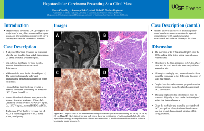
Case Description/Methods: A 63-year-old woman presented for neurosurgical evaluation after she was found to have a skull base mass on computerized tomography (CT) of the head at an outside hospital. She endorsed dysphagia for three months, however denied headaches or visual disturbances. A magnetic resonance imaging (MRI) revealed a 5.4 cm by 2.9 cm by 3.6 cm mass in the clivus, which was deemed as the cause of dysphagia (Figure 1a). The patient subsequently underwent an endoscopic transsphenoidal resection of the clival mass. Histopathology from the tissue revealed a hepatoid carcinoma, concerning for metastatic HCC (Figure 1b and 2c). Immunohistochemical strains were positive for hepatocytic marker arginase-1 (Figure 1d). Laboratory studies revealed alpha fetoprotein (AFP) of 56,344 ng/mL, CA-125 of 376 ng/mL, normal B-HCG and carcinoembryonic antigen (CEA). Thereafter, a triple phase CT of the liver revealed two LI-RADS 5 lesions suggestive of HCC as the primary malignancy. Patient’s case was discussed at multidisciplinary tumor board with recommendations for systemic immunotherapy with atezolimumab plus bevacizumab and radiation therapy to the clivus.
Discussion: The incidence of HCC has almost tripled since the 1980s making it the fastest rising cause of cancer related deaths. Metastasis to the brain comprises 0.26% to 2.2% of cases and the skull base is the most rarely affected anatomical site. Although CNS presentation is rare, we may see more neurological manifestations of metastatic HCC with the persistence of chronic hepatitis infections, the rise of metabolic diseases such as NASH, and an increase in alcohol-related liver disease during the COVID-19 pandemic. Although exceedingly rare, metastasis to the clivus should be considered in the differential diagnosis of skull base masses. Despite detection and treatment, prognosis remains poor and emphasis should be placed on consistent HCC surveillance. This case emphasizes that skull masses must be evaluated diligently as they can be the first sign of underlying liver malignancy. Given the morbidity and mortality associated with HCC, recognition of atypical manifestations of HCC can lead to a prompt diagnosis and initiation of life-saving treatment.

Disclosures:
Hunza Chaudhry, MD1, Sundeep Bekal, MD2, Alakh Gulati, MD2, Hongqi Peng, MD3, Marina Roytman, MD2. C0569 - Hepatocellular Carcinoma Presenting as a Clival Mass, ACG 2022 Annual Scientific Meeting Abstracts. Charlotte, NC: American College of Gastroenterology.
1UCSF-Fresno, Fresno, CA; 2UCSF Fresno, Fresno, CA; 3Community Regional Medical Center, Fresno, CA
Introduction: Hepatocellular carcinoma (HCC) comprises the majority of primary liver cancer and has a poor prognosis. Clivus metastasis is rare with only a few reported cases in the medical literature. We report a case of a patient who presented with clival mass found to have metastatic HCC.
Case Description/Methods: A 63-year-old woman presented for neurosurgical evaluation after she was found to have a skull base mass on computerized tomography (CT) of the head at an outside hospital. She endorsed dysphagia for three months, however denied headaches or visual disturbances. A magnetic resonance imaging (MRI) revealed a 5.4 cm by 2.9 cm by 3.6 cm mass in the clivus, which was deemed as the cause of dysphagia (Figure 1a). The patient subsequently underwent an endoscopic transsphenoidal resection of the clival mass. Histopathology from the tissue revealed a hepatoid carcinoma, concerning for metastatic HCC (Figure 1b and 2c). Immunohistochemical strains were positive for hepatocytic marker arginase-1 (Figure 1d). Laboratory studies revealed alpha fetoprotein (AFP) of 56,344 ng/mL, CA-125 of 376 ng/mL, normal B-HCG and carcinoembryonic antigen (CEA). Thereafter, a triple phase CT of the liver revealed two LI-RADS 5 lesions suggestive of HCC as the primary malignancy. Patient’s case was discussed at multidisciplinary tumor board with recommendations for systemic immunotherapy with atezolimumab plus bevacizumab and radiation therapy to the clivus.
Discussion: The incidence of HCC has almost tripled since the 1980s making it the fastest rising cause of cancer related deaths. Metastasis to the brain comprises 0.26% to 2.2% of cases and the skull base is the most rarely affected anatomical site. Although CNS presentation is rare, we may see more neurological manifestations of metastatic HCC with the persistence of chronic hepatitis infections, the rise of metabolic diseases such as NASH, and an increase in alcohol-related liver disease during the COVID-19 pandemic. Although exceedingly rare, metastasis to the clivus should be considered in the differential diagnosis of skull base masses. Despite detection and treatment, prognosis remains poor and emphasis should be placed on consistent HCC surveillance. This case emphasizes that skull masses must be evaluated diligently as they can be the first sign of underlying liver malignancy. Given the morbidity and mortality associated with HCC, recognition of atypical manifestations of HCC can lead to a prompt diagnosis and initiation of life-saving treatment.

Figure: Figure 1: A. Sagittal view of the MRI brain revealing clivus mass (red arrow). B. H&E stain at high power showing proliferation of malignant epithelial cells with hepatoid morphology arranged in sheets of nests and trabeculae. D. Positive immunohistochemical stain for hepatocytic marker arginase-1.
Disclosures:
Hunza Chaudhry indicated no relevant financial relationships.
Sundeep Bekal indicated no relevant financial relationships.
Alakh Gulati indicated no relevant financial relationships.
Hongqi Peng indicated no relevant financial relationships.
Marina Roytman indicated no relevant financial relationships.
Hunza Chaudhry, MD1, Sundeep Bekal, MD2, Alakh Gulati, MD2, Hongqi Peng, MD3, Marina Roytman, MD2. C0569 - Hepatocellular Carcinoma Presenting as a Clival Mass, ACG 2022 Annual Scientific Meeting Abstracts. Charlotte, NC: American College of Gastroenterology.
