Back


Poster Session A - Sunday Afternoon
Category: Biliary/Pancreas
A0045 - A Rare Pancreatic Tail Schwannoma in an Asymptomatic 58-Year-Old Female
Sunday, October 23, 2022
5:00 PM – 7:00 PM ET
Location: Crown Ballroom

Has Audio

Pranay Reddy, MD, MPH
Jefferson Health Northeast
Philadelphia, PA
Presenting Author(s)
Pranay Reddy, MD, MPH1, David Valadez, MD2, Mojtaba Olyaee, MD, FACG3
1Jefferson Health Northeast, Philadelphia, PA; 2University of Kansas Medical Center, Kansas City, KS; 3University of Kansas Medical Center, Leawood, KS
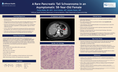
Introduction: Schwannomas are tumors which originate from Schwann cells responsible for fabricating myelin. Although Schwannomas are the most common benign peripheral nerve tumor in adults, there are several variants which are remarkably less common. Pancreatic schwannomas are an exceedingly rare type of nerve sheath tumor which arise from either sympathetic or parasympathetic vagal nerve fibers within the pancreas. In 2017, only 68 cases of pancreatic schwannoma had been reported in the preceding forty years with most occurring in the pancreatic head and body. In this case, we discuss an extraordinarily uncommon presentation of a pancreatic tail schwannoma in an asymptomatic 58-year-old female.
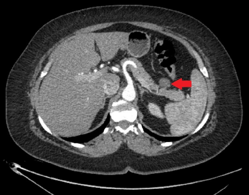
Case Description/Methods: A 58-year-old female with a past medical history of hypertension and hypothyroidism presented with findings of a 2 cm exophytic pancreatic tail lesion seen on prior CT imaging. The patient reportedly had a strong family history of aortic aneurysms and was found to have a right renal lesion on screening CT. She subsequently underwent CT abdomen and pelvis which revealed a lesion concerning for pancreatic tail malignancy. Endoscopic ultrasound (EUS) was performed which showed a 17 x 20 mm isoechoic peripheral pancreatic tail lesion. Fine needle aspiration (FNA) was performed which revealed spindle cells concerning for malignancy. Pathology and immunohistochemistry were inconclusive due to scant FNA aspirate obtained during EUS. Patient was taken to the operating room for exploratory laparotomy with distal pancreatectomy and splenectomy. Pathology of resected pancreatic mass showed typical histology with nuclear palisading and thick-walled vessels. Immunohistochemical staining supported the diagnosis of Schwannoma with diffuse, strong positivity for S-100 and SOX10 as well as negative staining for desmin, smooth muscle actin, CD34, pancytokeratin, CD117 and DOG1.
Discussion: Pancreatic schwannoma most commonly presents with abdominal pain although 30% of cases are found in asymptomatic patients with lesions discovered incidentally on screening CT scans. Although these lesions rarely display malignant transformation, they pose a significant diagnostic dilemma despite advances in radiographic imaging modalities. Endoscopic ultrasound is often limited by insufficient specimen collection and the preoperative diagnosis often becomes quite difficult. Enucleation of tumor is typically a sufficient therapeutic modality however radical resection is often required to establish the definitive diagnosis.

Disclosures:
Pranay Reddy, MD, MPH1, David Valadez, MD2, Mojtaba Olyaee, MD, FACG3. A0045 - A Rare Pancreatic Tail Schwannoma in an Asymptomatic 58-Year-Old Female, ACG 2022 Annual Scientific Meeting Abstracts. Charlotte, NC: American College of Gastroenterology.
1Jefferson Health Northeast, Philadelphia, PA; 2University of Kansas Medical Center, Kansas City, KS; 3University of Kansas Medical Center, Leawood, KS
Introduction: Schwannomas are tumors which originate from Schwann cells responsible for fabricating myelin. Although Schwannomas are the most common benign peripheral nerve tumor in adults, there are several variants which are remarkably less common. Pancreatic schwannomas are an exceedingly rare type of nerve sheath tumor which arise from either sympathetic or parasympathetic vagal nerve fibers within the pancreas. In 2017, only 68 cases of pancreatic schwannoma had been reported in the preceding forty years with most occurring in the pancreatic head and body. In this case, we discuss an extraordinarily uncommon presentation of a pancreatic tail schwannoma in an asymptomatic 58-year-old female.
Case Description/Methods: A 58-year-old female with a past medical history of hypertension and hypothyroidism presented with findings of a 2 cm exophytic pancreatic tail lesion seen on prior CT imaging. The patient reportedly had a strong family history of aortic aneurysms and was found to have a right renal lesion on screening CT. She subsequently underwent CT abdomen and pelvis which revealed a lesion concerning for pancreatic tail malignancy. Endoscopic ultrasound (EUS) was performed which showed a 17 x 20 mm isoechoic peripheral pancreatic tail lesion. Fine needle aspiration (FNA) was performed which revealed spindle cells concerning for malignancy. Pathology and immunohistochemistry were inconclusive due to scant FNA aspirate obtained during EUS. Patient was taken to the operating room for exploratory laparotomy with distal pancreatectomy and splenectomy. Pathology of resected pancreatic mass showed typical histology with nuclear palisading and thick-walled vessels. Immunohistochemical staining supported the diagnosis of Schwannoma with diffuse, strong positivity for S-100 and SOX10 as well as negative staining for desmin, smooth muscle actin, CD34, pancytokeratin, CD117 and DOG1.
Discussion: Pancreatic schwannoma most commonly presents with abdominal pain although 30% of cases are found in asymptomatic patients with lesions discovered incidentally on screening CT scans. Although these lesions rarely display malignant transformation, they pose a significant diagnostic dilemma despite advances in radiographic imaging modalities. Endoscopic ultrasound is often limited by insufficient specimen collection and the preoperative diagnosis often becomes quite difficult. Enucleation of tumor is typically a sufficient therapeutic modality however radical resection is often required to establish the definitive diagnosis.

Figure: CT scan showing 2 cm exophytic pancreatic tail lesion (red arrow)
Disclosures:
Pranay Reddy indicated no relevant financial relationships.
David Valadez indicated no relevant financial relationships.
Mojtaba Olyaee indicated no relevant financial relationships.
Pranay Reddy, MD, MPH1, David Valadez, MD2, Mojtaba Olyaee, MD, FACG3. A0045 - A Rare Pancreatic Tail Schwannoma in an Asymptomatic 58-Year-Old Female, ACG 2022 Annual Scientific Meeting Abstracts. Charlotte, NC: American College of Gastroenterology.
