Back


Poster Session B - Monday Morning
Category: General Endoscopy
B0299 - AL Amyloidosis Presenting as Isolated Gastric Lesion Found on Endoscopy in Patient With Recurrent Emesis
Monday, October 24, 2022
10:00 AM – 12:00 PM ET
Location: Crown Ballroom

Has Audio

Sara Gottesman, MD
Dell Medical School at the University of Texas at Austin
Austin, TX
Presenting Author(s)
Sara Gottesman, MD, David Ben-Nun, MD, Edgar Torres Fernandez, MD, Nirupama Ancha, , Kavitha Kumbum, MD
Dell Medical School at the University of Texas at Austin, Austin, TX
Introduction: Isolated gastric amyloid is a rare condition with a variable presentation. The following is a case of gastric amyloid presenting as a single large lesion in a patient with few nonspecific symptoms.
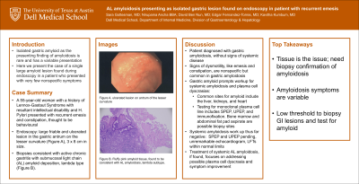
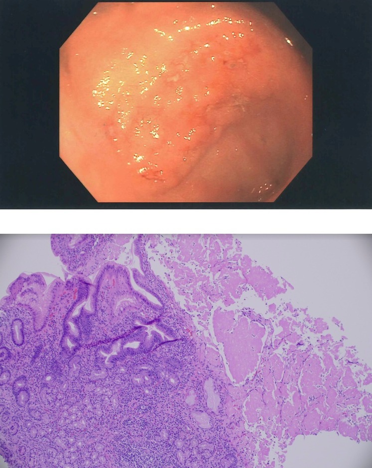
Case Description/Methods: A 55-year-old woman with a history of Lennox-Gastaut Syndrome underwent endoscopy for emesis workup. The procedure showed a large, friable, ulcerated lesion in the gastric antrum on the lesser curvature (Fig. A). The lesion measured approximately 3 x 8 cm and was concerning for malignancy. However, biopsies of the lesion were consistent with active chronic gastritis with submucosal light chain (AL) amyloid deposition, lambda type (Fig. B).
Discussion: Gastric amyloidosis is a rare condition, occurring in approximately only 3% of patients with amyloidosis. In light chain amyloidosis, it most often manifests in patients with the lambda subtype, as in the case presented here. The symptoms are vague and protean, making a clinical diagnosis difficult. Patients may complain of epigastric discomfort and may demonstrate signs of dysmotility. Ultimately, the definitive diagnosis and classification of amyloid must be made with histologic confirmation. In this case, the patient was unable to communicate her symptoms because of her intellectual disability, but did have constipation and increased emesis in the absence of other reported symptoms or findings consistent with systemic amyloidosis. Extensive workup to exclude systemic amyloidosis was unrevealing. This particular case is notable for the presentation of the disease with a single, large, friable ulcer, adding to the literature concerning the variable presentation of gastric amyloidosis on endoscopy. Considering the difficulty in clinical diagnosis and the morbidity of this disease, the authors propose a low threshold to consider pathologic examination for gastric amyloidosis in patients with nonspecific symptoms and abnormal findings on endoscopy.
Cowan, Skinner, et al. Amyloidosis of the gastrointestinal tract: a 13-year, single-center, referral experience. 2013. Haematologica., 98(1), 141–146.
Freudenthaler, S., et al. Amyloid in biopsies of the gastrointestinal tract-a retrospective observational study on 542 patients. 2016.Virchows Archiv : an international journal of pathology, 468(5), 569–577.
Said, S. et al.. Gastric amyloidosis: clinicopathological correlations in 79 cases from a single institution. 2015. Human pathology, 46(4), 491–498.

Disclosures:
Sara Gottesman, MD, David Ben-Nun, MD, Edgar Torres Fernandez, MD, Nirupama Ancha, , Kavitha Kumbum, MD. B0299 - AL Amyloidosis Presenting as Isolated Gastric Lesion Found on Endoscopy in Patient With Recurrent Emesis, ACG 2022 Annual Scientific Meeting Abstracts. Charlotte, NC: American College of Gastroenterology.
Dell Medical School at the University of Texas at Austin, Austin, TX
Introduction: Isolated gastric amyloid is a rare condition with a variable presentation. The following is a case of gastric amyloid presenting as a single large lesion in a patient with few nonspecific symptoms.
Case Description/Methods: A 55-year-old woman with a history of Lennox-Gastaut Syndrome underwent endoscopy for emesis workup. The procedure showed a large, friable, ulcerated lesion in the gastric antrum on the lesser curvature (Fig. A). The lesion measured approximately 3 x 8 cm and was concerning for malignancy. However, biopsies of the lesion were consistent with active chronic gastritis with submucosal light chain (AL) amyloid deposition, lambda type (Fig. B).
Discussion: Gastric amyloidosis is a rare condition, occurring in approximately only 3% of patients with amyloidosis. In light chain amyloidosis, it most often manifests in patients with the lambda subtype, as in the case presented here. The symptoms are vague and protean, making a clinical diagnosis difficult. Patients may complain of epigastric discomfort and may demonstrate signs of dysmotility. Ultimately, the definitive diagnosis and classification of amyloid must be made with histologic confirmation. In this case, the patient was unable to communicate her symptoms because of her intellectual disability, but did have constipation and increased emesis in the absence of other reported symptoms or findings consistent with systemic amyloidosis. Extensive workup to exclude systemic amyloidosis was unrevealing. This particular case is notable for the presentation of the disease with a single, large, friable ulcer, adding to the literature concerning the variable presentation of gastric amyloidosis on endoscopy. Considering the difficulty in clinical diagnosis and the morbidity of this disease, the authors propose a low threshold to consider pathologic examination for gastric amyloidosis in patients with nonspecific symptoms and abnormal findings on endoscopy.
Cowan, Skinner, et al. Amyloidosis of the gastrointestinal tract: a 13-year, single-center, referral experience. 2013. Haematologica., 98(1), 141–146.
Freudenthaler, S., et al. Amyloid in biopsies of the gastrointestinal tract-a retrospective observational study on 542 patients. 2016.Virchows Archiv : an international journal of pathology, 468(5), 569–577.
Said, S. et al.. Gastric amyloidosis: clinicopathological correlations in 79 cases from a single institution. 2015. Human pathology, 46(4), 491–498.

Figure: Fig A: This image was taken from endoscopy and shows a large friable, ulcerated lesion that was found in the patient's antrum on the lesser curvature. It was later found to be consistent with AL amyloidosis.
Fig. B: This is a histology slide from the biopsy that was taken from the gastric ulcer, demonstrating fluffy pink amyloid tissue, found to be consistent with AL amyloidosis, lambda subtype.
Fig. B: This is a histology slide from the biopsy that was taken from the gastric ulcer, demonstrating fluffy pink amyloid tissue, found to be consistent with AL amyloidosis, lambda subtype.
Disclosures:
Sara Gottesman indicated no relevant financial relationships.
David Ben-Nun indicated no relevant financial relationships.
Edgar Torres Fernandez indicated no relevant financial relationships.
Nirupama Ancha indicated no relevant financial relationships.
Kavitha Kumbum indicated no relevant financial relationships.
Sara Gottesman, MD, David Ben-Nun, MD, Edgar Torres Fernandez, MD, Nirupama Ancha, , Kavitha Kumbum, MD. B0299 - AL Amyloidosis Presenting as Isolated Gastric Lesion Found on Endoscopy in Patient With Recurrent Emesis, ACG 2022 Annual Scientific Meeting Abstracts. Charlotte, NC: American College of Gastroenterology.

