Back


Poster Session C - Monday Afternoon
Category: Small Intestine
C0664 - Hepatic Parenchyma and Cholecystectomy Clip in Duodenum? An Interesting Case of Pancreatic Adenocarcinoma
Monday, October 24, 2022
3:00 PM – 5:00 PM ET
Location: Crown Ballroom

Has Audio

Syed Musa Raza, MD
Louisiana State University Health Sciences Center
Shreveport, LA
Presenting Author(s)
Syed Musa Raza, MD1, Kristie R. Searcy, MD1, Jordan Roussel, MD1, Daniyal Raza, MD2, Rimsha Shaukat, MD1, Maryam Mubashir, MD1, Shazia Rashid, MD3, James Morris, MD, FACG4
1Louisiana State University Health Sciences Center, Shreveport, LA; 2Louisiana State University, Shreveport, LA; 3LSUHSC, Shreveport, LA; 4LSU Health Sciences Center, Shreveport, LA
Introduction: Pancreatic ductal adenocarcinoma (PDAC) is a leading cause of cancer-related death in many industrialized countries. Patients with PDAC typically report non-specific symptoms such as abdominal pain, weight loss, and jaundice. Given the lack of effective screening, many patients are diagnosed at the time of metastasis. Here we present a case of a patient who was diagnosed with metastatic PDAC with contained perforation in the duodenum.
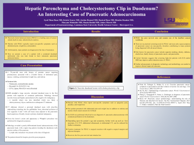
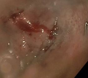
Case Description/Methods: A 74-year-old man with a history of prostate cancer requiring prostatectomy presented with a 2-month history of abdominal pain, nausea, vomiting, unintentional weight loss, and melena. He had normocytic and normochromic anemia with normal liver enzymes and lipase. Tumor markers ordered around the same time as EGD, including CEA/PSA, were unremarkable, but CA 19-9 was 6959 U/mL. EGD revealed a large necrotic, ulcerated duodenal mass in the first portion with suspicion of contained perforation. A metallic object was also identified which was likely a cholecystectomy clip as confirmed on subsequent CT. Pathology showed necrotic tissue and hepatic parenchymal cells, but there were no diagnostic neoplastic elements. CT abdomen showed a proximal duodenal mass with ulcer-like outpouching extending into the gallbladder fossa, abnormal perfusion of liver segments 4 and 5 with soft tissue mass adjacent to the pancreatic head suspicious of locally invasive primary duodenal malignancy. Whipple's procedure was performed, given the lesion's appearance and extent, and for diagnosis. Pathology revealed a poorly differentiated ductal adenocarcinoma of the head of the pancreas, with a signet ring pattern invading the duodenum wall, anterior surface of the pancreas. There was lymph node metastasis. The patient elected for hospice, given his poor prognosis.
Discussion: Patients with PDAC often report epigastric pain, jaundice, and weight loss. Our patient presented with abdominal pain and weight loss. However, the melena and anemia raised concern for GI bleed; hence an EGD was performed, which led to the diagnosis of pancreatic adenocarcinoma with contained perforation in the duodenum. There are no practical screening tools for pancreatic cancer. Liver enzymes, CA 19-9, in addition to ultrasound abdomen or abdominal CT, depending upon the patient's symptoms and age, can be considered as part of work up. Curative treatment for PDAC is surgical resection with negative surgical margins and adjuvant therapies; however, the five-year survival rate remains low.

Disclosures:
Syed Musa Raza, MD1, Kristie R. Searcy, MD1, Jordan Roussel, MD1, Daniyal Raza, MD2, Rimsha Shaukat, MD1, Maryam Mubashir, MD1, Shazia Rashid, MD3, James Morris, MD, FACG4. C0664 - Hepatic Parenchyma and Cholecystectomy Clip in Duodenum? An Interesting Case of Pancreatic Adenocarcinoma, ACG 2022 Annual Scientific Meeting Abstracts. Charlotte, NC: American College of Gastroenterology.
1Louisiana State University Health Sciences Center, Shreveport, LA; 2Louisiana State University, Shreveport, LA; 3LSUHSC, Shreveport, LA; 4LSU Health Sciences Center, Shreveport, LA
Introduction: Pancreatic ductal adenocarcinoma (PDAC) is a leading cause of cancer-related death in many industrialized countries. Patients with PDAC typically report non-specific symptoms such as abdominal pain, weight loss, and jaundice. Given the lack of effective screening, many patients are diagnosed at the time of metastasis. Here we present a case of a patient who was diagnosed with metastatic PDAC with contained perforation in the duodenum.
Case Description/Methods: A 74-year-old man with a history of prostate cancer requiring prostatectomy presented with a 2-month history of abdominal pain, nausea, vomiting, unintentional weight loss, and melena. He had normocytic and normochromic anemia with normal liver enzymes and lipase. Tumor markers ordered around the same time as EGD, including CEA/PSA, were unremarkable, but CA 19-9 was 6959 U/mL. EGD revealed a large necrotic, ulcerated duodenal mass in the first portion with suspicion of contained perforation. A metallic object was also identified which was likely a cholecystectomy clip as confirmed on subsequent CT. Pathology showed necrotic tissue and hepatic parenchymal cells, but there were no diagnostic neoplastic elements. CT abdomen showed a proximal duodenal mass with ulcer-like outpouching extending into the gallbladder fossa, abnormal perfusion of liver segments 4 and 5 with soft tissue mass adjacent to the pancreatic head suspicious of locally invasive primary duodenal malignancy. Whipple's procedure was performed, given the lesion's appearance and extent, and for diagnosis. Pathology revealed a poorly differentiated ductal adenocarcinoma of the head of the pancreas, with a signet ring pattern invading the duodenum wall, anterior surface of the pancreas. There was lymph node metastasis. The patient elected for hospice, given his poor prognosis.
Discussion: Patients with PDAC often report epigastric pain, jaundice, and weight loss. Our patient presented with abdominal pain and weight loss. However, the melena and anemia raised concern for GI bleed; hence an EGD was performed, which led to the diagnosis of pancreatic adenocarcinoma with contained perforation in the duodenum. There are no practical screening tools for pancreatic cancer. Liver enzymes, CA 19-9, in addition to ultrasound abdomen or abdominal CT, depending upon the patient's symptoms and age, can be considered as part of work up. Curative treatment for PDAC is surgical resection with negative surgical margins and adjuvant therapies; however, the five-year survival rate remains low.

Figure: Necrotic duodenal lesion with cholecystectomy clip
Disclosures:
Syed Musa Raza indicated no relevant financial relationships.
Kristie Searcy indicated no relevant financial relationships.
Jordan Roussel indicated no relevant financial relationships.
Daniyal Raza indicated no relevant financial relationships.
Rimsha Shaukat indicated no relevant financial relationships.
Maryam Mubashir indicated no relevant financial relationships.
Shazia Rashid indicated no relevant financial relationships.
James Morris: Celgene – Grant/Research Support.
Syed Musa Raza, MD1, Kristie R. Searcy, MD1, Jordan Roussel, MD1, Daniyal Raza, MD2, Rimsha Shaukat, MD1, Maryam Mubashir, MD1, Shazia Rashid, MD3, James Morris, MD, FACG4. C0664 - Hepatic Parenchyma and Cholecystectomy Clip in Duodenum? An Interesting Case of Pancreatic Adenocarcinoma, ACG 2022 Annual Scientific Meeting Abstracts. Charlotte, NC: American College of Gastroenterology.
