Back


Poster Session C - Monday Afternoon
Category: GI Bleeding
C0335 - Primary GI Mucosal Melanoma: A Rare Etiology of Iron Deficiency Anemia
Monday, October 24, 2022
3:00 PM – 5:00 PM ET
Location: Crown Ballroom

Has Audio

Usama Abu-Heija, MBBS
East Tennessee State University
Johnson City, TN
Presenting Author(s)
Usama Abu-Heija, MBBS, Mohammad Darweesh, MD, Damir Kusmic, MD, Mark Young, MD
East Tennessee State University, Johnson City, TN
Introduction: Iron deficiency anemia (IDA) is an alarming finding in males and post-menopausal females. A myriad of etiologies could be responsible for the anemia. In evaluation for possible GI blood loss, bidirectional endoscopy is usually warranted to identify a culprit for the anemia. We present a rare etiology of IDA identified on endoscopy in an elderly patient presenting with symptomatic anemia.
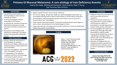
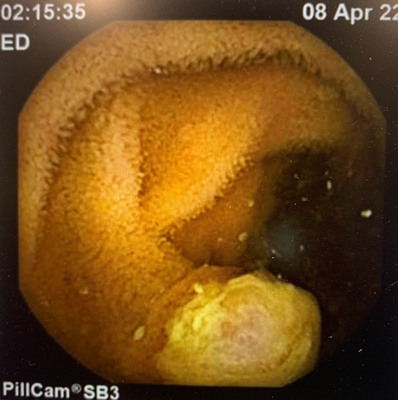
Case Description/Methods: An 89-year-old female with multiple medical comorbidities including atrial fibrillation on apixaban presented with fatigue, weakness, and exertional dyspnea. Laboratory investigations revealed IDA with a hemoglobin of 8.0 g/dl. Patient denied any overt GI bleeding. She was admitted, and a CT-Angiography of the abdomen revealed a questionable small focus of hemorrhage in the posterior gastric antrum, she was started on IV PPI and after adequate transfusion she underwent bidirectional endoscopy. Endoscopy findings were significant for a 3-4 mm raised umbilicated gastric lesion that was removed with cold forceps. The biopsy revealed a mucosal melanoma, SOX 10, Mart 1, HMB45 and S100 positive. Subsequently, the patient underwent a PET scan for staging which showed focal uptake at a distal esophageal lymph node; and focal uptake associated with a loop of small intestine in the left aspect of the pelvis. She also underwent a complete dermatological, ophthalmological evaluation and a Head CT scan to assess for a primary source, which were nonrevealing. To further evaluate the PET uptake in the small intestine she underwent video capsule endoscopy which revealed 3 small intestinal (likely jejunal) masses (figure 1) with a similar mucosal pattern to the identified gastric lesion. Patient was referred to Oncology and eventually underwent 3 rounds of radiotherapy followed by systemic chemotherapy.
Discussion: Malignant melanoma involving the GI tract can be either primary or metastatic. Primary GI mucosal melanoma is a rare entity with an annual incidence of 0.58 cases per million, its presentation is variable depending on location of the primary melanoma. Our case highlights the importance of performing a thorough endoscopic examination to identify subtle mucosal abnormalities that can aid in establishing an etiology of IDA in patients with occult GI bleeding who are on anticoagulation. Evaluation of malignant GI tract melanoma includes evaluation for a possible primary cutaneous source, as well as evaluating for metastatic disease.

Disclosures:
Usama Abu-Heija, MBBS, Mohammad Darweesh, MD, Damir Kusmic, MD, Mark Young, MD. C0335 - Primary GI Mucosal Melanoma: A Rare Etiology of Iron Deficiency Anemia, ACG 2022 Annual Scientific Meeting Abstracts. Charlotte, NC: American College of Gastroenterology.
East Tennessee State University, Johnson City, TN
Introduction: Iron deficiency anemia (IDA) is an alarming finding in males and post-menopausal females. A myriad of etiologies could be responsible for the anemia. In evaluation for possible GI blood loss, bidirectional endoscopy is usually warranted to identify a culprit for the anemia. We present a rare etiology of IDA identified on endoscopy in an elderly patient presenting with symptomatic anemia.
Case Description/Methods: An 89-year-old female with multiple medical comorbidities including atrial fibrillation on apixaban presented with fatigue, weakness, and exertional dyspnea. Laboratory investigations revealed IDA with a hemoglobin of 8.0 g/dl. Patient denied any overt GI bleeding. She was admitted, and a CT-Angiography of the abdomen revealed a questionable small focus of hemorrhage in the posterior gastric antrum, she was started on IV PPI and after adequate transfusion she underwent bidirectional endoscopy. Endoscopy findings were significant for a 3-4 mm raised umbilicated gastric lesion that was removed with cold forceps. The biopsy revealed a mucosal melanoma, SOX 10, Mart 1, HMB45 and S100 positive. Subsequently, the patient underwent a PET scan for staging which showed focal uptake at a distal esophageal lymph node; and focal uptake associated with a loop of small intestine in the left aspect of the pelvis. She also underwent a complete dermatological, ophthalmological evaluation and a Head CT scan to assess for a primary source, which were nonrevealing. To further evaluate the PET uptake in the small intestine she underwent video capsule endoscopy which revealed 3 small intestinal (likely jejunal) masses (figure 1) with a similar mucosal pattern to the identified gastric lesion. Patient was referred to Oncology and eventually underwent 3 rounds of radiotherapy followed by systemic chemotherapy.
Discussion: Malignant melanoma involving the GI tract can be either primary or metastatic. Primary GI mucosal melanoma is a rare entity with an annual incidence of 0.58 cases per million, its presentation is variable depending on location of the primary melanoma. Our case highlights the importance of performing a thorough endoscopic examination to identify subtle mucosal abnormalities that can aid in establishing an etiology of IDA in patients with occult GI bleeding who are on anticoagulation. Evaluation of malignant GI tract melanoma includes evaluation for a possible primary cutaneous source, as well as evaluating for metastatic disease.

Figure: Figure 1
Disclosures:
Usama Abu-Heija indicated no relevant financial relationships.
Mohammad Darweesh indicated no relevant financial relationships.
Damir Kusmic indicated no relevant financial relationships.
Mark Young indicated no relevant financial relationships.
Usama Abu-Heija, MBBS, Mohammad Darweesh, MD, Damir Kusmic, MD, Mark Young, MD. C0335 - Primary GI Mucosal Melanoma: A Rare Etiology of Iron Deficiency Anemia, ACG 2022 Annual Scientific Meeting Abstracts. Charlotte, NC: American College of Gastroenterology.
