Poster Session C - Monday Afternoon
Category: Stomach
C0686 - Metastatic Gastric Cancer to the Rectum: A Systematic Review


Dawood Findakly, MD
Feist-Weiller Cancer Center, Louisiana State University Health
Shreveport, LA
Presenting Author(s)
Feist-Weiller Cancer Center, Louisiana State University Health, Shreveport, LA
Introduction: Rectal metastasis (RM) from gastric cancer (GC) is an extremely rare entity. Herein, we review trends to promote detection and strategies for management.
Methods: PubMed search for "gastric cancer" and "rectal metastasis," from database inception through April 30, 2022; pooled with a case from our institution.
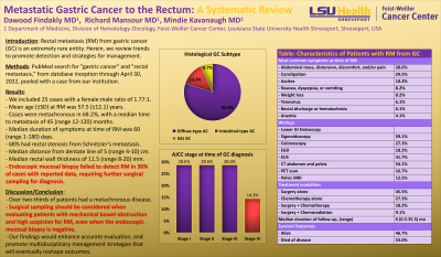
Results: We included 25 cases with a female:male ratio of 1.77:1. Mean age (±SD) at RM was 57.5 (±12.1) years. Cases were metachronous in 68.2%, with a median time to RM of 45 (range 12-120) months. Histologically, 80% had diffuse-type adenocarcinoma (AC), 13.3% had intestinal-type AC, and 6.7% had gastroesophageal junction AC originating from the stomach.
Regarding initial GC diagnosis (iGCd) AJCC stage and TNM classification, 28.6% were stage (St IIA, T3N0M0), the rest were evenly split at 14.3% for each of St IA (T1N0M0), St IB (T2N0M0), St IIIA (T3N2M0), St IIIB, and St IV (T3N0M1). Treatment included surgery alone (45.5%), chemotherapy (27.3%), surgery with chemotherapy (18.2%), and surgery with chemoradiation (9.1%). Rectal stenosis from Schnitzler's metastasis described in 68% of cases. The most common symptoms at RM were mechanical bowel obstruction—including abdominal mass, distension, discomfort, and/or pain (28.6%); constipation (24.5%); ascites (10.2%); and nausea, dyspepsia, or vomiting (8.2%), weight loss (8.2%), tenesmus (6.1%), rectal discharge or hematochezia (6.1%), and anemia (4.1%). RM median duration of symptoms was 60 (range 1-180) days with a median distance from dentate line of 5 (range 4-10) cm and a median rectal wall thickness of 11.5 (range 8-20) mm.
All patients underwent lower endoscopy—sigmoidoscopy in 59.1%, and colonoscopy in 27.3%. EUS in 41.7%, CT abdomen/pelvis in 54.1%, EGD in 18.2%—75% were synchronous AC. Pelvic MRI was utilized in 12.5%. PET scan was utilized in 16.7%—all had established GC diagnosis, 75% presented with rectal stenosis, and 75% failed to detect RM on endoscopic mucosal biopsy requiring further surgical sampling.
Regarding outcomes, 46.7% were alive and 53.0% succumbed to disease with a median follow-up of 6 (range 0.5-35.5) months after RM.
Discussion: In our cohort, over two-thirds of patients had a metachronous disease. Surgical sampling should be performed to detect RM when evaluating patients with mechanical bowel obstruction, even when the endoscopic mucosal biopsy is negative. Our findings would enhance accurate evaluation, and promote multidisciplinary management strategies that will eventually reshape outcomes.
Characteristics of Patients with RM from GC | |
Female:Male | 1.77:1 |
Mean age, (±SD) | 57.5, (±12.1) years |
Metachronous | 68.2% |
- Median time to RM in metachronous setting, (range) | 45, (12-120) months |
Histological subtype | |
- Diffuse type AC | 80% |
- Intestinal type AC | 13.3% |
- Gastroesophageal junction originating from the stomach | 6.7% |
AJCC stage groups at the time of GC diagnosis, (TNM) | |
- St IA (T1N0M0) | 14.3% |
- St IB (T2N0M0) | 14.3% |
- St IIA (T3N0M0) | 28.6% |
- St IIIA (T3N2M0) | 14.3% |
- St IIIB (NM) | 14.3% |
- St IV (T3N0M1) | 14.3% |
Rectal stenosis from Schnitzler's metastasis | 68% |
Most common symptoms at time of RM | |
- Abdominal mass, distension, discomfort, and/or pain | 28.6% |
- Constipation | 24.5% |
- Ascites | 10.2% |
- Nausea, dyspepsia, or vomiting | 8.2% |
- Weight loss | 8.2% |
- Tenesmus | 6.1% |
- Rectal discharge or hematochezia | 6.1% |
- Anemia | 4.1% |
Median duration of symptoms at the time of RM diagnosis, (range) | 60, (1-180) days |
Median distance of RM from the dentate line, (range) | 5, (4-10) cm |
Median rectal wall thickness, (range) | 11.5 (8-20) mm |
Workup | |
- Lower GI Endoscopy |
|
- Sigmoidoscopy | 59.1% |
- Colonoscopy | 27.3% |
- EGD | 18.2% |
- EUS | 41.7% |
- CT abdomen and pelvis | 54.1% |
- PET scan | 16.7% |
- Pelvic MRI | 12.5% |
Median duration of follow-up, (range) | 6 (0.5-35.5) months |
Treatment modalities |
|
- S alone | 45.5% |
- C alone | 27.3% |
- S+C | 18.2% |
- S+CRT | 9.1% |
Survival Outcomes |
|
- Alive | 46.7% |
- Died of disease | 53.0% |
Legend: RM: rectal metastasis; GC: gastric cancer; AC: adenocarcinoma; AJCC: American Joint Committee on Cancer; St: stage; TNM: TNM classification of malignant tumors; NM: not mentioned; S: surgery; C: chemotherapy; CRT: chemoradiation; GI: gastrointestinal; EGD: esophagogastroduodenoscopy; EUS: endoscopic ultrasound; CT: computed tomography; PET: positron emission tomography; MRI: magnetic resonance imaging. | |
Disclosures:
Dawood Findakly, MD, Richard P. Mansour, MD, Mindie Kavanaugh, MD. C0686 - Metastatic Gastric Cancer to the Rectum: A Systematic Review, ACG 2022 Annual Scientific Meeting Abstracts. Charlotte, NC: American College of Gastroenterology.
