Back


Poster Session B - Monday Morning
Category: General Endoscopy
B0297 - The Xiphisternum as a Gastric Subepithelial Lesion
Monday, October 24, 2022
10:00 AM – 12:00 PM ET
Location: Crown Ballroom

Has Audio

Aoife Feighery, MBBCh
Mayo Clinic
Rochester, MN
Presenting Author(s)
Aoife Feighery, MBBCh1, David Prichard, MBBCh, BAO, PhD2
1Mayo Clinic, Rochester, MN; 2Trinity College Dublin, Dublin, Dublin, Ireland
Introduction: The xiphisternum, comprised of cartilage surrounding a core of bone, is located inferior to the sternal body and enlarges with age. The process is usually directed anteriorly relative to the sternal body and the abdominal cavity. However, in approximately 10% of individuals the bony structure is angulated more than 10 degrees posterior to the orientation of the sternal body. Although this anatomical variant is typically asymptomatic, it may be encountered during endoscopy. As such, endoscopists should be familiar with its appearance to ensure correct identification.
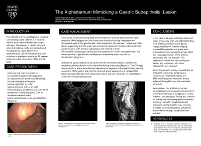
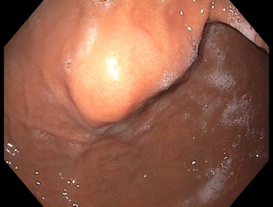
Case Description/Methods: A 66-year-old man presented to an outpatient gastroenterology clinic for assessment of diarrhea and bloating. He had a background medical history significant for colon adenocarcinoma with prior right hemicolectomy, prostate cancer, and B Cell lymphoma. During diagnostic EGD, an incidental medium-sized gastric subepithelial lesion was identified (Figure 1). Biopsy of the lesion demonstrated gastric mucosa with foveolar hyperplasia and minimal chronic inflammation. Endoscopic ultrasound was obtained for further characterization. The lesion was visualized causing indentation on the anterior wall of the gastric body. No mucosal abnormalities were present. With respiration, the stomach “rolled over” this lesion, suggesting that the origin was extramural. Endosonography demonstrated a hyperechoic, multilayered, shadowing lesion external to the stomach. Palpation of the epigastrium resulted in indentation of the stomach just below the lesion. A review of sagittal sections of an abdominal CT, performed for unrelated reasons, clarified the underlying etiology for the lesion identified during endoscopy.
Discussion: The CT image demonstrates a posteriorly directed xiphisternum adjacent to the gastric body, causing protrusion of the gastric wall into the stomach which appeared as a subepithelial lesion during endoscopy. Upon visualizing this structure for this patient specifically, it was important to consider lymphoma, or compressive lymphadenopathy, as a differential diagnosis. However, with an intraprocedural examination and information available at the time of the EGD, it is likely that the endoscopic ultrasound could have been avoided. This was likely an incidental finding and not related to his presenting diarrhea and so no intervention was pursued.

Disclosures:
Aoife Feighery, MBBCh1, David Prichard, MBBCh, BAO, PhD2. B0297 - The Xiphisternum as a Gastric Subepithelial Lesion, ACG 2022 Annual Scientific Meeting Abstracts. Charlotte, NC: American College of Gastroenterology.
1Mayo Clinic, Rochester, MN; 2Trinity College Dublin, Dublin, Dublin, Ireland
Introduction: The xiphisternum, comprised of cartilage surrounding a core of bone, is located inferior to the sternal body and enlarges with age. The process is usually directed anteriorly relative to the sternal body and the abdominal cavity. However, in approximately 10% of individuals the bony structure is angulated more than 10 degrees posterior to the orientation of the sternal body. Although this anatomical variant is typically asymptomatic, it may be encountered during endoscopy. As such, endoscopists should be familiar with its appearance to ensure correct identification.
Case Description/Methods: A 66-year-old man presented to an outpatient gastroenterology clinic for assessment of diarrhea and bloating. He had a background medical history significant for colon adenocarcinoma with prior right hemicolectomy, prostate cancer, and B Cell lymphoma. During diagnostic EGD, an incidental medium-sized gastric subepithelial lesion was identified (Figure 1). Biopsy of the lesion demonstrated gastric mucosa with foveolar hyperplasia and minimal chronic inflammation. Endoscopic ultrasound was obtained for further characterization. The lesion was visualized causing indentation on the anterior wall of the gastric body. No mucosal abnormalities were present. With respiration, the stomach “rolled over” this lesion, suggesting that the origin was extramural. Endosonography demonstrated a hyperechoic, multilayered, shadowing lesion external to the stomach. Palpation of the epigastrium resulted in indentation of the stomach just below the lesion. A review of sagittal sections of an abdominal CT, performed for unrelated reasons, clarified the underlying etiology for the lesion identified during endoscopy.
Discussion: The CT image demonstrates a posteriorly directed xiphisternum adjacent to the gastric body, causing protrusion of the gastric wall into the stomach which appeared as a subepithelial lesion during endoscopy. Upon visualizing this structure for this patient specifically, it was important to consider lymphoma, or compressive lymphadenopathy, as a differential diagnosis. However, with an intraprocedural examination and information available at the time of the EGD, it is likely that the endoscopic ultrasound could have been avoided. This was likely an incidental finding and not related to his presenting diarrhea and so no intervention was pursued.

Figure: Figure 1: Subepithelial gastric lesion identified during endoscopy
Disclosures:
Aoife Feighery indicated no relevant financial relationships.
David Prichard indicated no relevant financial relationships.
Aoife Feighery, MBBCh1, David Prichard, MBBCh, BAO, PhD2. B0297 - The Xiphisternum as a Gastric Subepithelial Lesion, ACG 2022 Annual Scientific Meeting Abstracts. Charlotte, NC: American College of Gastroenterology.
