Back


Poster Session B - Monday Morning
Category: Interventional Endoscopy
B0462 - Comparison of EGD to EGD With EUS in Evaluation of Sub-Epithelial Lesions of the Upper GI Tract
Monday, October 24, 2022
10:00 AM – 12:00 PM ET
Location: Crown Ballroom

Has Audio
- WB
William Bigelow, MD
University of Virginia Health System
Charlottesville, VA
Presenting Author(s)
William Bigelow, MD1, Vanessa M. Shami, MD, FACG2, Bryan Sauer, MD, FACG2, Ross Buerlein, MD3
1University of Virginia Health System, Charlottesville, VA; 2University of Virginia Digestive Health Center, Charlottesville, VA; 3University of Virginia Digestive Health Center, Keswick, VA
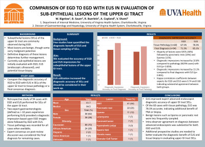
Introduction: Subepithelial lesions of the upper GI tract are commonly observed during esophagogastroduodenoscopy (EGD). While mostly benign, definitive diagnosis of these lesions can determine further management. Currently sub-epithelial lesions are initially evaluated with EGD, EUS (endoscopic ultrasound), and tissue biopsy. In this study, we compared the diagnostic yield of subepithelial lesions with EGD alone to EGD with EUS.
Methods: 94 cases of subepithelial lesions performed at an academic tertiary referral medical center containing both EGD and EUS reports with images were examined retrospectively. Two expert gastroenterologists (defined as greater than ten years experience performing EGD and EUS) reviewed each case separately and recorded a diagnostic impression based upon EGD alone followed by an impression of EGD with EUS. Tissue pathology was recorded for all cases when available. For cases without definitive tissue pathology, expert consensus on post-review discussion was considered the final diagnosis.
Results: Expert impressions of subepithelial lesions on EGD alone and EGD with EUS were 71.3% and 85.1% accurate respectively. A 13.8% increase in diagnostic accuracy was observed with the addition of EUS (p=0.001). There were 60 cases with definitive tissue pathology available. In these cases, EGD alone and EGD with EUS impressions were 67.5% and 78.3% accurate respectively in comparison to final pathology. A 10.8% increase in diagnostic accuracy was observed with addition of EUS (p=0.059). Expert agreement occurred in 78.7% of EGD alone impressions and 88.3% of EGD with EUS impressions.
Discussion: Additional evaluation of subepithelial lesions with EUS characterization demonstrated an increase in diagnostic accuracy compared to EGD alone. These results illustrate the utility of EUS in diagnosis of these potentially malignant lesions. Future studies should continue to evaluate the diagnostic benefit from both EUS and various biopsy techniques to further develop guideline-driven approaches in evaluation of subepithelial lesions.
Disclosures:
William Bigelow, MD1, Vanessa M. Shami, MD, FACG2, Bryan Sauer, MD, FACG2, Ross Buerlein, MD3. B0462 - Comparison of EGD to EGD With EUS in Evaluation of Sub-Epithelial Lesions of the Upper GI Tract, ACG 2022 Annual Scientific Meeting Abstracts. Charlotte, NC: American College of Gastroenterology.
1University of Virginia Health System, Charlottesville, VA; 2University of Virginia Digestive Health Center, Charlottesville, VA; 3University of Virginia Digestive Health Center, Keswick, VA
Introduction: Subepithelial lesions of the upper GI tract are commonly observed during esophagogastroduodenoscopy (EGD). While mostly benign, definitive diagnosis of these lesions can determine further management. Currently sub-epithelial lesions are initially evaluated with EGD, EUS (endoscopic ultrasound), and tissue biopsy. In this study, we compared the diagnostic yield of subepithelial lesions with EGD alone to EGD with EUS.
Methods: 94 cases of subepithelial lesions performed at an academic tertiary referral medical center containing both EGD and EUS reports with images were examined retrospectively. Two expert gastroenterologists (defined as greater than ten years experience performing EGD and EUS) reviewed each case separately and recorded a diagnostic impression based upon EGD alone followed by an impression of EGD with EUS. Tissue pathology was recorded for all cases when available. For cases without definitive tissue pathology, expert consensus on post-review discussion was considered the final diagnosis.
Results: Expert impressions of subepithelial lesions on EGD alone and EGD with EUS were 71.3% and 85.1% accurate respectively. A 13.8% increase in diagnostic accuracy was observed with the addition of EUS (p=0.001). There were 60 cases with definitive tissue pathology available. In these cases, EGD alone and EGD with EUS impressions were 67.5% and 78.3% accurate respectively in comparison to final pathology. A 10.8% increase in diagnostic accuracy was observed with addition of EUS (p=0.059). Expert agreement occurred in 78.7% of EGD alone impressions and 88.3% of EGD with EUS impressions.
Discussion: Additional evaluation of subepithelial lesions with EUS characterization demonstrated an increase in diagnostic accuracy compared to EGD alone. These results illustrate the utility of EUS in diagnosis of these potentially malignant lesions. Future studies should continue to evaluate the diagnostic benefit from both EUS and various biopsy techniques to further develop guideline-driven approaches in evaluation of subepithelial lesions.
Disclosures:
William Bigelow indicated no relevant financial relationships.
Vanessa Shami: Boston Scientific – Giving a lecture funded by Boston Scientific at ACG interventional fellows course. cook medical – Consultant. olympus – Consultant.
Bryan Sauer indicated no relevant financial relationships.
Ross Buerlein indicated no relevant financial relationships.
William Bigelow, MD1, Vanessa M. Shami, MD, FACG2, Bryan Sauer, MD, FACG2, Ross Buerlein, MD3. B0462 - Comparison of EGD to EGD With EUS in Evaluation of Sub-Epithelial Lesions of the Upper GI Tract, ACG 2022 Annual Scientific Meeting Abstracts. Charlotte, NC: American College of Gastroenterology.
