Back


Poster Session B - Monday Morning
Category: General Endoscopy
B0295 - Combined Endoscopic Ultrasound and Endobronchial Ultrasound to Stage and Diagnose Non-Small Cell Lung Cancer in the Setting of CLL/SLL
Monday, October 24, 2022
10:00 AM – 12:00 PM ET
Location: Crown Ballroom

Has Audio
- WG
William C. Green, MD
Wake Forest University School of Medicine
Winston-Salem, NC
Presenting Author(s)
William C. Green, MD1, Kathleen Herring, MD2, Cynthia Ezedike, MD3, Jason Conway, MD4
1Wake Forest University School of Medicine, Winston-Salem, NC; 2Internal Medicine Residency Program, Wake Forest University School of Medicine, Winston-Salem, NC; 3Wake Forest Baptist Health, Winston-Salem, NC; 4Wake Forest Baptist, Winston-Salem, NC
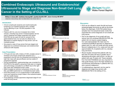
Introduction: Chronic lymphocytic leukemia and small lymphocytic lymphoma is an indolent leukemia with a variable course affecting 4-5 per 100,000 population in the US/year. Patients with CLL are at an increased risk of other cancers, including lung cancer. Endosonography can be used to diagnose and stage suspected lung cancer. EUS-FNA can be used not only for cancers of the gastrointestinal tract, but can also be utilized as an alternative to surgical staging for lung cancer, such as mediastinoscopy. We present a case of lung cancer that was staged and diagnosed via combined endoscopic and endobronchial ultrasound with biopsies.
Case Description/Methods: A 78 year-old man with a history of CAD, prostate cancer in remission and chronic lymphocytic leukemia (CLL) presented to the ED from his scheduled bronchoscopy for a right hilar mass with pleural effusion and two weeks of progressive dyspnea. During hospitalization, gastroenterology was consulted for a EUS-FNA for the patient’s subcarinal node seen on imaging and pulmonology consulted for an EBUS due to this left hilar mass with mediastinal lymph nodes. EBUS revealed an extrinsic compression of the RLL and RUL and EBNA performed on 3 mediastinal lymph nodes. EUS-FNA showed 3-4 cm conglomeration of well-defined, round and hypoechoic lymph nodes in the subcarinal space with fine needle biopsy. Final pathology of these specimens diagnosed stage III non-small cell lung cancer.
Discussion: EUS can be utilized to reach the left and lower paraesophageal structures as well as structures below the diaphragm. EUS-FNA added tissue samples from his subcarinal lymph nodes that supported the correct diagnosis of non-small cell lung cancer. Due to the diagnosis of non-small cell lung cancer from his combined EBUS/EUS-FNA, the patient did not require surgical staging.
Our case demonstrates the need use of a combined EUS/EBUS approach to diagnose a patient with CLL with non-small cell lung cancer. Second malignancies in CLL cases are rare and approximately 2% of patients with CLL develop lung carcinoma. Recent studies showed that patients with CLL less than 55 years old have a greater risk of developing secondary solid tumors, not like our patient who was 78 years old. Tissue samples from the EUS-FNA/EBUS are very important to help guide therapy due to the high resistance to conventional chemotherapy in patients with CLL and a second malignancy.
A multidisciplinary approach can be used to diagnose and stage lung cancers with EBUS/EUS-FNA in the setting of CLL.
Disclosures:
William C. Green, MD1, Kathleen Herring, MD2, Cynthia Ezedike, MD3, Jason Conway, MD4. B0295 - Combined Endoscopic Ultrasound and Endobronchial Ultrasound to Stage and Diagnose Non-Small Cell Lung Cancer in the Setting of CLL/SLL, ACG 2022 Annual Scientific Meeting Abstracts. Charlotte, NC: American College of Gastroenterology.
1Wake Forest University School of Medicine, Winston-Salem, NC; 2Internal Medicine Residency Program, Wake Forest University School of Medicine, Winston-Salem, NC; 3Wake Forest Baptist Health, Winston-Salem, NC; 4Wake Forest Baptist, Winston-Salem, NC
Introduction: Chronic lymphocytic leukemia and small lymphocytic lymphoma is an indolent leukemia with a variable course affecting 4-5 per 100,000 population in the US/year. Patients with CLL are at an increased risk of other cancers, including lung cancer. Endosonography can be used to diagnose and stage suspected lung cancer. EUS-FNA can be used not only for cancers of the gastrointestinal tract, but can also be utilized as an alternative to surgical staging for lung cancer, such as mediastinoscopy. We present a case of lung cancer that was staged and diagnosed via combined endoscopic and endobronchial ultrasound with biopsies.
Case Description/Methods: A 78 year-old man with a history of CAD, prostate cancer in remission and chronic lymphocytic leukemia (CLL) presented to the ED from his scheduled bronchoscopy for a right hilar mass with pleural effusion and two weeks of progressive dyspnea. During hospitalization, gastroenterology was consulted for a EUS-FNA for the patient’s subcarinal node seen on imaging and pulmonology consulted for an EBUS due to this left hilar mass with mediastinal lymph nodes. EBUS revealed an extrinsic compression of the RLL and RUL and EBNA performed on 3 mediastinal lymph nodes. EUS-FNA showed 3-4 cm conglomeration of well-defined, round and hypoechoic lymph nodes in the subcarinal space with fine needle biopsy. Final pathology of these specimens diagnosed stage III non-small cell lung cancer.
Discussion: EUS can be utilized to reach the left and lower paraesophageal structures as well as structures below the diaphragm. EUS-FNA added tissue samples from his subcarinal lymph nodes that supported the correct diagnosis of non-small cell lung cancer. Due to the diagnosis of non-small cell lung cancer from his combined EBUS/EUS-FNA, the patient did not require surgical staging.
Our case demonstrates the need use of a combined EUS/EBUS approach to diagnose a patient with CLL with non-small cell lung cancer. Second malignancies in CLL cases are rare and approximately 2% of patients with CLL develop lung carcinoma. Recent studies showed that patients with CLL less than 55 years old have a greater risk of developing secondary solid tumors, not like our patient who was 78 years old. Tissue samples from the EUS-FNA/EBUS are very important to help guide therapy due to the high resistance to conventional chemotherapy in patients with CLL and a second malignancy.
A multidisciplinary approach can be used to diagnose and stage lung cancers with EBUS/EUS-FNA in the setting of CLL.
Disclosures:
William Green indicated no relevant financial relationships.
Kathleen Herring indicated no relevant financial relationships.
Cynthia Ezedike indicated no relevant financial relationships.
Jason Conway: Cook Medical – Consultant.
William C. Green, MD1, Kathleen Herring, MD2, Cynthia Ezedike, MD3, Jason Conway, MD4. B0295 - Combined Endoscopic Ultrasound and Endobronchial Ultrasound to Stage and Diagnose Non-Small Cell Lung Cancer in the Setting of CLL/SLL, ACG 2022 Annual Scientific Meeting Abstracts. Charlotte, NC: American College of Gastroenterology.
