Back


Poster Session C - Monday Afternoon
Category: Esophagus
C0232 - Esophageal Hemorrhage After Transesophageal Echocardiography in a Patient With Undiagnosed Eosinophilic Esophagitis
Monday, October 24, 2022
3:00 PM – 5:00 PM ET
Location: Crown Ballroom

Has Audio

John T. Nathanson, MD
New York-Presbyterian/Columbia University Medical Center
New York, NY
Presenting Author(s)
John T. Nathanson, MD1, Shawn Chawla, MD1, John Blackett, MD2, Reuben J. Garcia-Carrasquillo, MD, FACG3
1New York-Presbyterian/Columbia University Medical Center, New York, NY; 2Mayo Clinic, Rochester, MN; 3Columbia University College of Physicians and Surgeons, New York, NY
Introduction: Eosinophilic esophagitis (EoE) is characterized by infiltration of the esophageal epithelium by eosinophils. The burden of disease is likely underreported, with a prevalence of 57 cases per 100,000. Patients experience gastroesophageal reflux, dysphagia, and recurrent food impactions, with spontaneous rupture being a rare but serious complication. Characteristic endoscopic findings include stacked circular rings, strictures, and linear furrows. Esophageal biopsy must demonstrate ≥ 15 eosinophils per high-power field. Transesophageal echocardiography (TEE) allows for detailed evaluation of cardiac structure and function and is performed via the blind passage of a probe into the esophagus and stomach. Patients with esophageal abnormalities are at increased risk for esophageal injury during TEE.
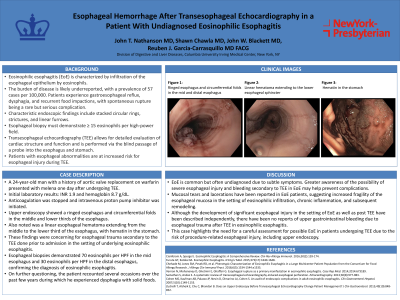
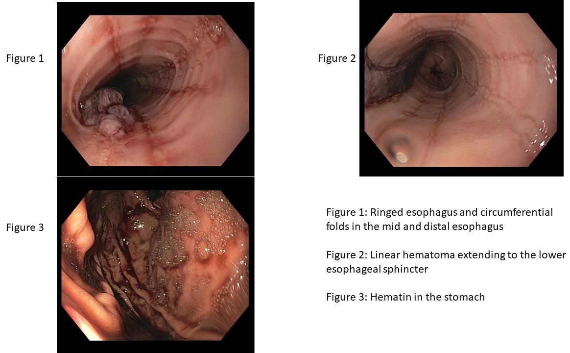
Case Description/Methods: A 24-year-old man with a history of aortic valve replacement on warfarin presented with melena one day after undergoing TEE. Initial laboratory results showed INR 1.9 and hemoglobin 8.7 g/dL. Anticoagulation was stopped and intravenous proton pump inhibitor was initiated. Upper endoscopy showed a ringed esophagus and circumferential folds in the middle and lower thirds of the esophagus. Also noted was a linear esophageal hematoma extending from the middle to the lower third of the esophagus, with hematin in the stomach. These findings were concerning for esophageal trauma secondary to the TEE done prior to admission in the setting of underlying eosinophilic esophagitis. Esophageal biopsies demonstrated 70 eosinophils per HPF in the mid esophagus and 30 eosinophils per HPF in the distal esophagus, confirming the diagnosis of eosinophilic esophagitis. On further questioning, the patient recounted several occasions over the past few years during which he experienced dysphagia with solid foods.
Discussion: Mucosal tears and lacerations have been reported in EoE patients, suggesting increased fragility of the esophageal mucosa in the setting of eosinophilic infiltration, chronic inflammation, and subsequent remodeling. Although the development of significant esophageal injury in the setting of EoE as well as post TEE have been described independently, there have been no reports of upper gastrointestinal bleeding due to esophageal trauma after TEE in eosinophilic esophagitis. This case highlights the need for a careful assessment for possible EoE in patients undergoing TEE due to the risk of procedure-related esophageal injury.

Disclosures:
John T. Nathanson, MD1, Shawn Chawla, MD1, John Blackett, MD2, Reuben J. Garcia-Carrasquillo, MD, FACG3. C0232 - Esophageal Hemorrhage After Transesophageal Echocardiography in a Patient With Undiagnosed Eosinophilic Esophagitis, ACG 2022 Annual Scientific Meeting Abstracts. Charlotte, NC: American College of Gastroenterology.
1New York-Presbyterian/Columbia University Medical Center, New York, NY; 2Mayo Clinic, Rochester, MN; 3Columbia University College of Physicians and Surgeons, New York, NY
Introduction: Eosinophilic esophagitis (EoE) is characterized by infiltration of the esophageal epithelium by eosinophils. The burden of disease is likely underreported, with a prevalence of 57 cases per 100,000. Patients experience gastroesophageal reflux, dysphagia, and recurrent food impactions, with spontaneous rupture being a rare but serious complication. Characteristic endoscopic findings include stacked circular rings, strictures, and linear furrows. Esophageal biopsy must demonstrate ≥ 15 eosinophils per high-power field. Transesophageal echocardiography (TEE) allows for detailed evaluation of cardiac structure and function and is performed via the blind passage of a probe into the esophagus and stomach. Patients with esophageal abnormalities are at increased risk for esophageal injury during TEE.
Case Description/Methods: A 24-year-old man with a history of aortic valve replacement on warfarin presented with melena one day after undergoing TEE. Initial laboratory results showed INR 1.9 and hemoglobin 8.7 g/dL. Anticoagulation was stopped and intravenous proton pump inhibitor was initiated. Upper endoscopy showed a ringed esophagus and circumferential folds in the middle and lower thirds of the esophagus. Also noted was a linear esophageal hematoma extending from the middle to the lower third of the esophagus, with hematin in the stomach. These findings were concerning for esophageal trauma secondary to the TEE done prior to admission in the setting of underlying eosinophilic esophagitis. Esophageal biopsies demonstrated 70 eosinophils per HPF in the mid esophagus and 30 eosinophils per HPF in the distal esophagus, confirming the diagnosis of eosinophilic esophagitis. On further questioning, the patient recounted several occasions over the past few years during which he experienced dysphagia with solid foods.
Discussion: Mucosal tears and lacerations have been reported in EoE patients, suggesting increased fragility of the esophageal mucosa in the setting of eosinophilic infiltration, chronic inflammation, and subsequent remodeling. Although the development of significant esophageal injury in the setting of EoE as well as post TEE have been described independently, there have been no reports of upper gastrointestinal bleeding due to esophageal trauma after TEE in eosinophilic esophagitis. This case highlights the need for a careful assessment for possible EoE in patients undergoing TEE due to the risk of procedure-related esophageal injury.

Figure: Figure 1: Ringed esophagus and circumferential folds in the mid and distal esophagus
Figure 2: Linear hematoma extending to the lower esophageal sphincter
Figure 3: Hematin in the stomach
Figure 2: Linear hematoma extending to the lower esophageal sphincter
Figure 3: Hematin in the stomach
Disclosures:
John Nathanson indicated no relevant financial relationships.
Shawn Chawla indicated no relevant financial relationships.
John Blackett indicated no relevant financial relationships.
Reuben Garcia-Carrasquillo indicated no relevant financial relationships.
John T. Nathanson, MD1, Shawn Chawla, MD1, John Blackett, MD2, Reuben J. Garcia-Carrasquillo, MD, FACG3. C0232 - Esophageal Hemorrhage After Transesophageal Echocardiography in a Patient With Undiagnosed Eosinophilic Esophagitis, ACG 2022 Annual Scientific Meeting Abstracts. Charlotte, NC: American College of Gastroenterology.
