Back


Poster Session B - Monday Morning
Category: Interventional Endoscopy
B0469 - Uterine Cervical Squamous Cell Carcinoma: A Rare Cause of Biliary Obstruction
Monday, October 24, 2022
10:00 AM – 12:00 PM ET
Location: Crown Ballroom

Has Audio
- AR
Aaron Rampersad, MD
Prisma Health
Greenville, SC
Presenting Author(s)
Aaron Rampersad, MD1, Elizabeth Martin, MD1, Kari Valente, MD1, Wesley Jones, BS, MD1, William Bolton, MD1, Veeral Oza, MD2
1Prisma Health, Greenville, SC; 2University of South Carolina School of Medicine Greenville, Prisma Health, Greenville, SC
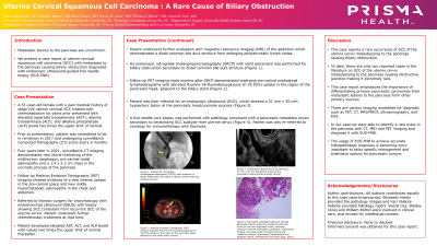
Introduction: The incidence of biliary obstruction is 5 cases per 1000 people in the United States with reports of 21% caused by metastatic lesions to the pancreaticobiliary system. Metastatic lesion to the pancreas is rare, with few cases on uterine squamous cell cancer (SCC) metastasizing to the pancreas. We present a case report of a uterine cervical SCC with metastasis to the pancreas resulting in biliary obstruction.
Case Description/Methods: A 51-year-old female with a history of stage 4b uterine cervical SCC previously treated with radiation and chemotherapy six years prior presented with elevated aspartate transaminase (AST), alanine transaminase (ALT), and alkaline phosphatase (ALP) levels two times the upper limit of normal and jaundice.
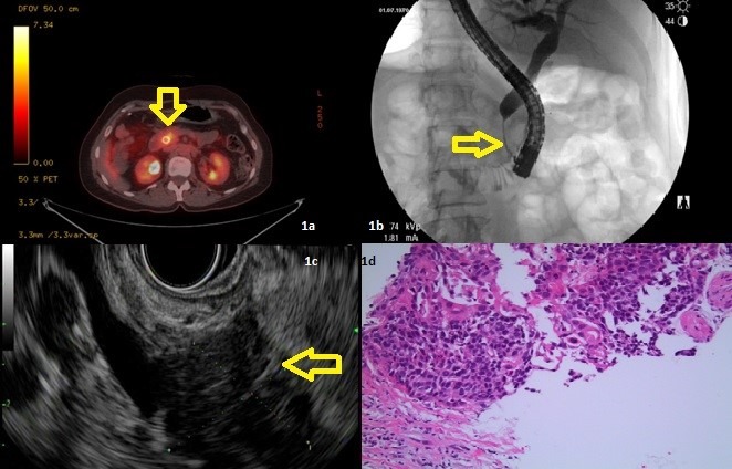
Prior to presentation she was in remission. However, CT on presentation demonstrated new thickening of the midthoracic esophagus, with subcarinal lymphadenopathy and a 1.9 x 2.2 cm mass in the uncinate process of the pancreas causing biliary dilation. Follow-up PET imaging showed evidence of a new intense uptake in the pre-carinal space and new pancreatic head involvement (Fig 1a).
She simultaneously underwent an Endoscopic Ultrasound (EUS) and Endoscopic retrograde cholangiopancreatography (ERCP) for stent placement (Fig 1b). EUS showed a 21 mm x 20 mm hypoechoic lesion at the pancreatic head/uncinate process (Figure 1c). A fine needle biopsy was performed. Pathologic evaluation was consistent with a metastatic lesion secondary to a keratinizing SCC subtype was identified. (Figure 1d).
Discussion: Metastasis to the pancreas accounts for 2-5% of pancreatic malignancies. Majority of SCC are associated with cervical high risk-human papilloma virus (HPV) with HPV-16 and HPV-18 being responsible for the most common types of cervical cancers. To date there have been only two published reports in the literature on SCC of the uterine cervix metastasizing to the pancreas. This case report emphasizes the importance of differentiating primary pancreatic carcinomas from metastatic lesions to the pancreas from other primary sources. There are various imaging modalities for diagnosis such as PET, CT, MRI/MRCP, ultrasonography, and EUS. In our case we were able to identify a new mass in the pancreas with CT, MRI and PET imaging and diagnose it with EUS. The usage of EUS to achieve accurate histopathologic diagnosis is becoming more important to tailor specific management and treatment options for pancreatic tumors

Disclosures:
Aaron Rampersad, MD1, Elizabeth Martin, MD1, Kari Valente, MD1, Wesley Jones, BS, MD1, William Bolton, MD1, Veeral Oza, MD2. B0469 - Uterine Cervical Squamous Cell Carcinoma: A Rare Cause of Biliary Obstruction, ACG 2022 Annual Scientific Meeting Abstracts. Charlotte, NC: American College of Gastroenterology.
1Prisma Health, Greenville, SC; 2University of South Carolina School of Medicine Greenville, Prisma Health, Greenville, SC
Introduction: The incidence of biliary obstruction is 5 cases per 1000 people in the United States with reports of 21% caused by metastatic lesions to the pancreaticobiliary system. Metastatic lesion to the pancreas is rare, with few cases on uterine squamous cell cancer (SCC) metastasizing to the pancreas. We present a case report of a uterine cervical SCC with metastasis to the pancreas resulting in biliary obstruction.
Case Description/Methods: A 51-year-old female with a history of stage 4b uterine cervical SCC previously treated with radiation and chemotherapy six years prior presented with elevated aspartate transaminase (AST), alanine transaminase (ALT), and alkaline phosphatase (ALP) levels two times the upper limit of normal and jaundice.
Prior to presentation she was in remission. However, CT on presentation demonstrated new thickening of the midthoracic esophagus, with subcarinal lymphadenopathy and a 1.9 x 2.2 cm mass in the uncinate process of the pancreas causing biliary dilation. Follow-up PET imaging showed evidence of a new intense uptake in the pre-carinal space and new pancreatic head involvement (Fig 1a).
She simultaneously underwent an Endoscopic Ultrasound (EUS) and Endoscopic retrograde cholangiopancreatography (ERCP) for stent placement (Fig 1b). EUS showed a 21 mm x 20 mm hypoechoic lesion at the pancreatic head/uncinate process (Figure 1c). A fine needle biopsy was performed. Pathologic evaluation was consistent with a metastatic lesion secondary to a keratinizing SCC subtype was identified. (Figure 1d).
Discussion: Metastasis to the pancreas accounts for 2-5% of pancreatic malignancies. Majority of SCC are associated with cervical high risk-human papilloma virus (HPV) with HPV-16 and HPV-18 being responsible for the most common types of cervical cancers. To date there have been only two published reports in the literature on SCC of the uterine cervix metastasizing to the pancreas. This case report emphasizes the importance of differentiating primary pancreatic carcinomas from metastatic lesions to the pancreas from other primary sources. There are various imaging modalities for diagnosis such as PET, CT, MRI/MRCP, ultrasonography, and EUS. In our case we were able to identify a new mass in the pancreas with CT, MRI and PET imaging and diagnose it with EUS. The usage of EUS to achieve accurate histopathologic diagnosis is becoming more important to tailor specific management and treatment options for pancreatic tumors

Figure: Fig 1a: PET avid lesion; 1b: Stricture as seen on ERCP within the distal CBD; 1c: Pancreatic mass on EUS; 1d: pancreatic tissue involved by sheets of cells with round to oval and angulated nuclei, moderate amounts of dense eosinophilic cytoplasm, intercellular bridges, and focal keratin formation. Scattered mitoses, dyskeratotic keratinocytes and apoptotic debris
Disclosures:
Aaron Rampersad indicated no relevant financial relationships.
Elizabeth Martin indicated no relevant financial relationships.
Kari Valente indicated no relevant financial relationships.
Wesley Jones indicated no relevant financial relationships.
William Bolton indicated no relevant financial relationships.
Veeral Oza: Boston Scientific – Consultant. S4 Medical – Advisor or Review Panel Member.
Aaron Rampersad, MD1, Elizabeth Martin, MD1, Kari Valente, MD1, Wesley Jones, BS, MD1, William Bolton, MD1, Veeral Oza, MD2. B0469 - Uterine Cervical Squamous Cell Carcinoma: A Rare Cause of Biliary Obstruction, ACG 2022 Annual Scientific Meeting Abstracts. Charlotte, NC: American College of Gastroenterology.
