Back


Poster Session D - Tuesday Morning
Category: Biliary/Pancreas
D0036 - A Rare Case of Symptomatic Lymphoepithelial Cyst of the Pancreas
Tuesday, October 25, 2022
10:00 AM – 12:00 PM ET
Location: Crown Ballroom

Has Audio

James Rock, DO
Allegheny Health Network
Pittsburgh, PA
Presenting Author(s)
James Rock, DO, Christina DiMaria, DO, Marcia Mitre, MD
Allegheny Health Network, Pittsburgh, PA
Introduction: Lymphoproliferative cysts (LEC) of the pancreas are rare, nonmalignant cysts. LECs of the pancreas are true pancreatic cysts as defined by their squamous epithelium lining, while being surrounded by mature lymphoid tissue. These cysts can be very difficult to differentiate from other pancreatic cysts or neoplasms on imaging. Patients with LEC cysts will typically present with nausea, abdominal pain, unintentional weight loss, vomiting, and diarrhea as the cyst grows. In 2013, there were 109 documented cases of LEC cysts in the literature (1). Here, we describe a patient presenting with a LEC of the pancreas.
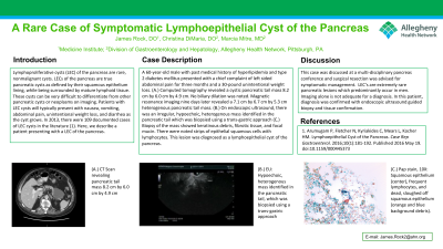
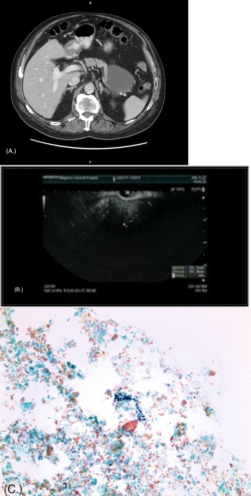
Case Description/Methods: A 60-year-old male with past medical history of hyperlipidemia and type 2 diabetes mellitus presented with a chief complaint of left sided abdominal pain for three months and a 30-pound unintentional weight loss. (A.) Computed tomography revealed a cystic pancreatic tail mass 8.2 cm by 6.0 cm by 4.9 cm. No biliary dilation was noted. Magnetic resonance imaging nine days later revealed a 7.1 cm by 6.7 cm by 5.3 cm heterogenous pancreatic tail mass. (B.) On endoscopic ultrasound, there was an irregular, hypoechoic, heterogenous mass identified in the pancreatic tail which was biopsied using a trans-gastric approach (C.) Biopsy of the mass showed keratinous debris, fibrotic tissue, and focal mucin. There were noted strips of epithelial squamous cells with lymphocytes. This lesion was diagnosed as a lymphoepithelial cyst of the pancreas.
Discussion: This case was discussed at a multi-disciplinary pancreas conference and surgical resection was advised for symptomatic management. LEC’s are extremely rare pancreatic lesions which predominantly occur in men. Imaging alone is not adequate for a diagnosis. In this patient, diagnosis was confirmed with endoscopic ultrasound guided biopsy.

Disclosures:
James Rock, DO, Christina DiMaria, DO, Marcia Mitre, MD. D0036 - A Rare Case of Symptomatic Lymphoepithelial Cyst of the Pancreas, ACG 2022 Annual Scientific Meeting Abstracts. Charlotte, NC: American College of Gastroenterology.
Allegheny Health Network, Pittsburgh, PA
Introduction: Lymphoproliferative cysts (LEC) of the pancreas are rare, nonmalignant cysts. LECs of the pancreas are true pancreatic cysts as defined by their squamous epithelium lining, while being surrounded by mature lymphoid tissue. These cysts can be very difficult to differentiate from other pancreatic cysts or neoplasms on imaging. Patients with LEC cysts will typically present with nausea, abdominal pain, unintentional weight loss, vomiting, and diarrhea as the cyst grows. In 2013, there were 109 documented cases of LEC cysts in the literature (1). Here, we describe a patient presenting with a LEC of the pancreas.
Case Description/Methods: A 60-year-old male with past medical history of hyperlipidemia and type 2 diabetes mellitus presented with a chief complaint of left sided abdominal pain for three months and a 30-pound unintentional weight loss. (A.) Computed tomography revealed a cystic pancreatic tail mass 8.2 cm by 6.0 cm by 4.9 cm. No biliary dilation was noted. Magnetic resonance imaging nine days later revealed a 7.1 cm by 6.7 cm by 5.3 cm heterogenous pancreatic tail mass. (B.) On endoscopic ultrasound, there was an irregular, hypoechoic, heterogenous mass identified in the pancreatic tail which was biopsied using a trans-gastric approach (C.) Biopsy of the mass showed keratinous debris, fibrotic tissue, and focal mucin. There were noted strips of epithelial squamous cells with lymphocytes. This lesion was diagnosed as a lymphoepithelial cyst of the pancreas.
Discussion: This case was discussed at a multi-disciplinary pancreas conference and surgical resection was advised for symptomatic management. LEC’s are extremely rare pancreatic lesions which predominantly occur in men. Imaging alone is not adequate for a diagnosis. In this patient, diagnosis was confirmed with endoscopic ultrasound guided biopsy.

Figure: A. CT Scan revealing pancreatic tail mass 8.2 cm by 6.0 cm by 4.9 cm
B. EU: Hypoechoic, heterogenous mass identified in the pancreatic tail, which was biopsied using a trans-gastric approach
C. Pap stain, 10X: Pap stain demonstrating squamous epithelium (center), frequent lymphocytes (small, dark, round cells throughout the field) and dead, sloughed off squamous epithelium (orange and blue background debris). The presence of squamous epithelium and lymphocytes on a fine-needle biopsy of the pancreas supports the diagnosis of pancreatic lymphoepithelial cyst
Disclosures:
James Rock indicated no relevant financial relationships.
Christina DiMaria indicated no relevant financial relationships.
Marcia Mitre indicated no relevant financial relationships.
James Rock, DO, Christina DiMaria, DO, Marcia Mitre, MD. D0036 - A Rare Case of Symptomatic Lymphoepithelial Cyst of the Pancreas, ACG 2022 Annual Scientific Meeting Abstracts. Charlotte, NC: American College of Gastroenterology.
