Back


Poster Session B - Monday Morning
Category: Colon
B0108 - The Unexpected Quiet Guest in the Colon
Monday, October 24, 2022
10:00 AM – 12:00 PM ET
Location: Crown Ballroom

Has Audio

Omar Tageldin, MD
Albany Medical Center
Menands, NY
Presenting Author(s)
Omar Tageldin, MD1, Spyridon Zouridis, MD1, Hala Abdelwahab, MBBCh1, Andrea Lightle, DO1, James J. Litynski, MD2
1Albany Medical Center, Albany, NY; 2Albany Medical Center, Niskayuna, NY
Introduction: Schistosomiasis is a common parasitic infection, more prevalent in Sub-Saharan Africa but rare in developed countries. Schistosomiasis is transmitted through skin contact with fresh water while snails serve as intermediate hosts. Other than intestinal and hepatosplenic schistosomiasis, pulmonary manifestations and genitourinary infection may develop. Gastrointestinal symptoms may include abdominal pain, nausea, bloody diarrhea, tenesmus, and hepatosplenomegaly. We present a case of completely asymptomatic intestinal schistosomiasis diagnosed incidentally during colonoscopy.
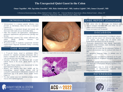
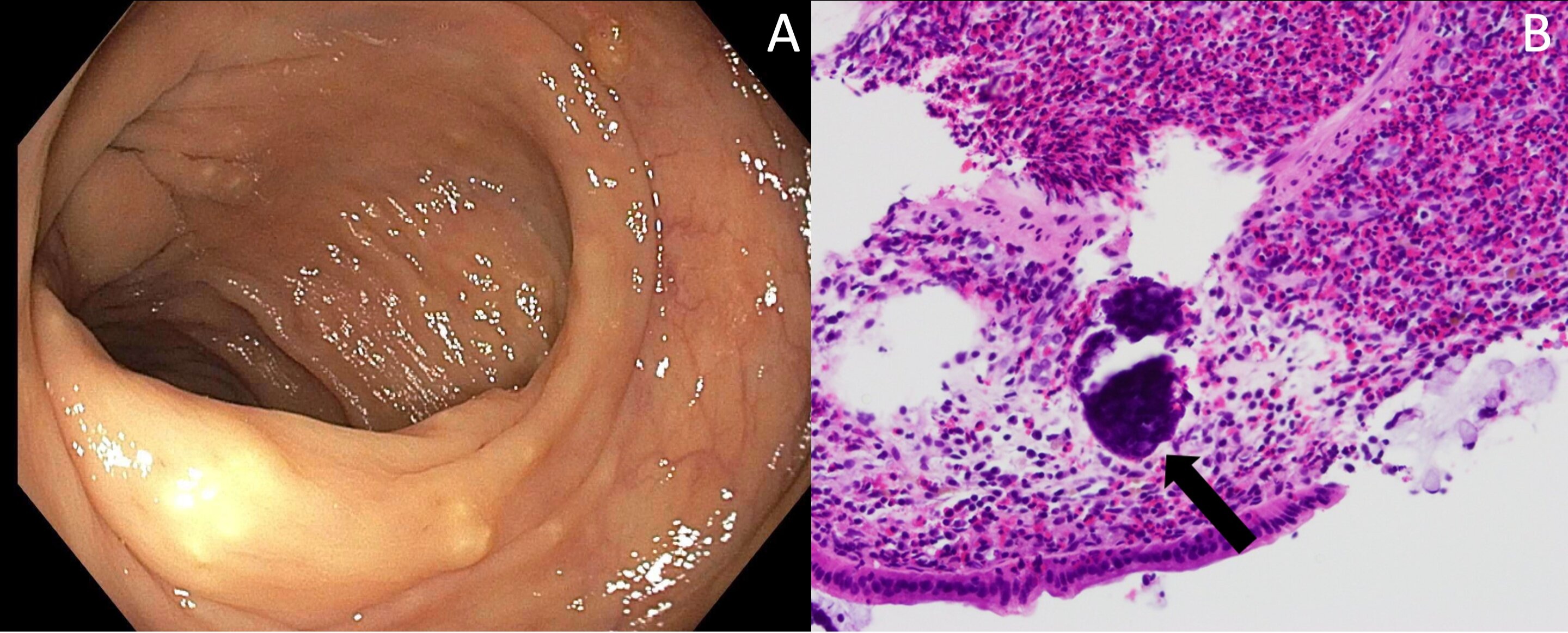
Case Description/Methods: A 57-year-old female, raised in sub-Saharan Africa, presented for outpatient colonoscopy after a positive Cologuard test, she didn’t have any complaints and her physical exam was benign. Colonoscopy was performed and revealed nodular mucosa in the cecum, splenic flexure, and the rectum. The splenic flexure nodules were characterized by a unique white tipped appearance [Figure 1.A]. Biopsies were obtained from these nodules and the histopathology showed colonic mucosa with an eosinophilic granulomatous inflammation associated with Schistosoma egg at the splenic flexure nodular lesions [Figure 1.B]. The other nodular areas were identified as benign colonic mucosa with lymphoid aggregate. Stool culture after the colonoscopy was obtained and showed incidental Endolimax nana cysts which was not treated as per infectious disease recommendations. Patient was treated by a course of Praziquantel.
Discussion: Typically, intestinal schistosomiasis is considered a chronic infection and may present with abdominal pain, diarrhea, poor appetite, and weight loss. In severe cases, hematochezia and anemia may also be observed. Endoscopic characteristics may include hypertrophy, granulomas, polyp formation, mural thickening, strictures with erosions or ulcerations. The endoscopic findings of intestinal schistosomiasis may be confused with those of IBD. For appropriate diagnosis, biopsy in multiple sites is required. Schistosoma eggs surrounded by granulomas are the gold standard for diagnosis. Peripheral eosinophilia also supports the diagnosis. Patients with intestinal schistosomiasis are treated with a single dose of Praziquantel 40mg/kg regardless of the symptoms. Gastroenterologists must be aware of the endoscopic characteristics of schistosomiasis, especially when managing patients from endemic areas.

Disclosures:
Omar Tageldin, MD1, Spyridon Zouridis, MD1, Hala Abdelwahab, MBBCh1, Andrea Lightle, DO1, James J. Litynski, MD2. B0108 - The Unexpected Quiet Guest in the Colon, ACG 2022 Annual Scientific Meeting Abstracts. Charlotte, NC: American College of Gastroenterology.
1Albany Medical Center, Albany, NY; 2Albany Medical Center, Niskayuna, NY
Introduction: Schistosomiasis is a common parasitic infection, more prevalent in Sub-Saharan Africa but rare in developed countries. Schistosomiasis is transmitted through skin contact with fresh water while snails serve as intermediate hosts. Other than intestinal and hepatosplenic schistosomiasis, pulmonary manifestations and genitourinary infection may develop. Gastrointestinal symptoms may include abdominal pain, nausea, bloody diarrhea, tenesmus, and hepatosplenomegaly. We present a case of completely asymptomatic intestinal schistosomiasis diagnosed incidentally during colonoscopy.
Case Description/Methods: A 57-year-old female, raised in sub-Saharan Africa, presented for outpatient colonoscopy after a positive Cologuard test, she didn’t have any complaints and her physical exam was benign. Colonoscopy was performed and revealed nodular mucosa in the cecum, splenic flexure, and the rectum. The splenic flexure nodules were characterized by a unique white tipped appearance [Figure 1.A]. Biopsies were obtained from these nodules and the histopathology showed colonic mucosa with an eosinophilic granulomatous inflammation associated with Schistosoma egg at the splenic flexure nodular lesions [Figure 1.B]. The other nodular areas were identified as benign colonic mucosa with lymphoid aggregate. Stool culture after the colonoscopy was obtained and showed incidental Endolimax nana cysts which was not treated as per infectious disease recommendations. Patient was treated by a course of Praziquantel.
Discussion: Typically, intestinal schistosomiasis is considered a chronic infection and may present with abdominal pain, diarrhea, poor appetite, and weight loss. In severe cases, hematochezia and anemia may also be observed. Endoscopic characteristics may include hypertrophy, granulomas, polyp formation, mural thickening, strictures with erosions or ulcerations. The endoscopic findings of intestinal schistosomiasis may be confused with those of IBD. For appropriate diagnosis, biopsy in multiple sites is required. Schistosoma eggs surrounded by granulomas are the gold standard for diagnosis. Peripheral eosinophilia also supports the diagnosis. Patients with intestinal schistosomiasis are treated with a single dose of Praziquantel 40mg/kg regardless of the symptoms. Gastroenterologists must be aware of the endoscopic characteristics of schistosomiasis, especially when managing patients from endemic areas.

Figure: Figure1. A. White tipped nodular mucosa at splenic flexure. B. H&E section showing colonic mucosa with an eosinophilic granulomatous inflammation associated with Schistosoma egg (arrow). [20x magnification].
Disclosures:
Omar Tageldin indicated no relevant financial relationships.
Spyridon Zouridis indicated no relevant financial relationships.
Hala Abdelwahab indicated no relevant financial relationships.
Andrea Lightle indicated no relevant financial relationships.
James Litynski indicated no relevant financial relationships.
Omar Tageldin, MD1, Spyridon Zouridis, MD1, Hala Abdelwahab, MBBCh1, Andrea Lightle, DO1, James J. Litynski, MD2. B0108 - The Unexpected Quiet Guest in the Colon, ACG 2022 Annual Scientific Meeting Abstracts. Charlotte, NC: American College of Gastroenterology.
