Back


Poster Session D - Tuesday Morning
Category: Biliary/Pancreas
D0060 - Hepatolithiasis Caused by Right Hepatic Artery Branches Forming an Arterial Ring Compressing the Common Hepatic Duct
Tuesday, October 25, 2022
10:00 AM – 12:00 PM ET
Location: Crown Ballroom

Has Audio

David A. Iannitti, Sr., MD
Atrium Health
Charlotte, NC
Presenting Author(s)
Award: Presidential Poster Award
David A. Iannitti, MD1, Yifan Wang, MD2, Erin Baker, MD1, Matt Strand, MD1, Stephen Deal, MD3
1Atrium Health, Charlotte, NC; 2McGill University, Charlotte, NC; 3Carolina Digestive, Charlotte, NC
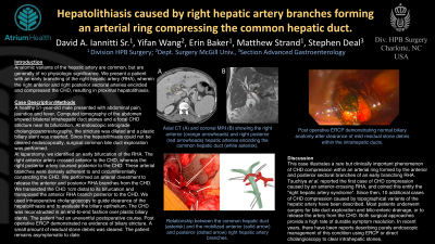
Introduction: Anatomic variants of the hepatic artery are common, but are generally of no physiologic significance. We present a patient with an early branching of the right hepatic artery (RHA), wherein the right anterior and right posterior sectoral arteries encircled and compressed the CHD, resulting in proximal hepatolithiasis.
Case Description/Methods: A healthy 51-year-old male presented with abdominal pain, jaundice and fever. Computed tomography of the abdomen showed bilateral intrahepatic duct stones and a focal CHD stricture near its bifurcation. At endoscopic retrograde cholangiopancreatography, the stricture was dilated and a plastic biliary stent was inserted. Since the hepatolithiasis could not be cleared endoscopically, surgical common bile duct exploration was performed.
At laparotomy, we identified an early bifurcation of the RHA. The right anterior artery crossed anterior to the CHD, whereas the right posterior artery coursed posterior to the CHD. These arterial branches were densely adherent to and circumferentially constricting the CHD. We performed an arterial divestment to release the anterior and posterior RHA branches from the CHD. We transected the CHD 1cm distal to its bifurcation and transposed the anterior RHA branch posterior to the CHD. We used Spyglass cholangioscopy to guide clearance of the hepatolithiasis and to evaluate the biliary epithelium. The CHD was reconstructed in an end-to-end fashion over plastic biliary stents. The patient had an uneventful postoperative course. Post operative ERCP demonstrated no evidence of stones or biliary stricture. The patient remains asymptomatic to date.
Discussion: This case illustrates a rare but clinically important phenomenon of CHD compression within an arterial ring formed by the anterior and posterior sectoral branches of an early branching RHA. Tsuchiya et al. reported the first case of CHD compression caused by an anterior-crossing RHA, and coined this entity the “right hepatic artery syndrome”. Since then, 10 additional cases of CHD compression caused by topographical variants of the hepatic artery have been described. Most patients underwent surgery for bile duct exploration and bilioenteric drainage, or to release the artery from the CHD. Both surgical approaches provide a high rate of durable symptom resolution. In recent years, there have been reports describing purely endoscopic management of this condition using ERCP or direct cholangioscopy to clear intrahepatic stones.

Disclosures:
David A. Iannitti, MD1, Yifan Wang, MD2, Erin Baker, MD1, Matt Strand, MD1, Stephen Deal, MD3. D0060 - Hepatolithiasis Caused by Right Hepatic Artery Branches Forming an Arterial Ring Compressing the Common Hepatic Duct, ACG 2022 Annual Scientific Meeting Abstracts. Charlotte, NC: American College of Gastroenterology.
David A. Iannitti, MD1, Yifan Wang, MD2, Erin Baker, MD1, Matt Strand, MD1, Stephen Deal, MD3
1Atrium Health, Charlotte, NC; 2McGill University, Charlotte, NC; 3Carolina Digestive, Charlotte, NC
Introduction: Anatomic variants of the hepatic artery are common, but are generally of no physiologic significance. We present a patient with an early branching of the right hepatic artery (RHA), wherein the right anterior and right posterior sectoral arteries encircled and compressed the CHD, resulting in proximal hepatolithiasis.
Case Description/Methods: A healthy 51-year-old male presented with abdominal pain, jaundice and fever. Computed tomography of the abdomen showed bilateral intrahepatic duct stones and a focal CHD stricture near its bifurcation. At endoscopic retrograde cholangiopancreatography, the stricture was dilated and a plastic biliary stent was inserted. Since the hepatolithiasis could not be cleared endoscopically, surgical common bile duct exploration was performed.
At laparotomy, we identified an early bifurcation of the RHA. The right anterior artery crossed anterior to the CHD, whereas the right posterior artery coursed posterior to the CHD. These arterial branches were densely adherent to and circumferentially constricting the CHD. We performed an arterial divestment to release the anterior and posterior RHA branches from the CHD. We transected the CHD 1cm distal to its bifurcation and transposed the anterior RHA branch posterior to the CHD. We used Spyglass cholangioscopy to guide clearance of the hepatolithiasis and to evaluate the biliary epithelium. The CHD was reconstructed in an end-to-end fashion over plastic biliary stents. The patient had an uneventful postoperative course. Post operative ERCP demonstrated no evidence of stones or biliary stricture. The patient remains asymptomatic to date.
Discussion: This case illustrates a rare but clinically important phenomenon of CHD compression within an arterial ring formed by the anterior and posterior sectoral branches of an early branching RHA. Tsuchiya et al. reported the first case of CHD compression caused by an anterior-crossing RHA, and coined this entity the “right hepatic artery syndrome”. Since then, 10 additional cases of CHD compression caused by topographical variants of the hepatic artery have been described. Most patients underwent surgery for bile duct exploration and bilioenteric drainage, or to release the artery from the CHD. Both surgical approaches provide a high rate of durable symptom resolution. In recent years, there have been reports describing purely endoscopic management of this condition using ERCP or direct cholangioscopy to clear intrahepatic stones.

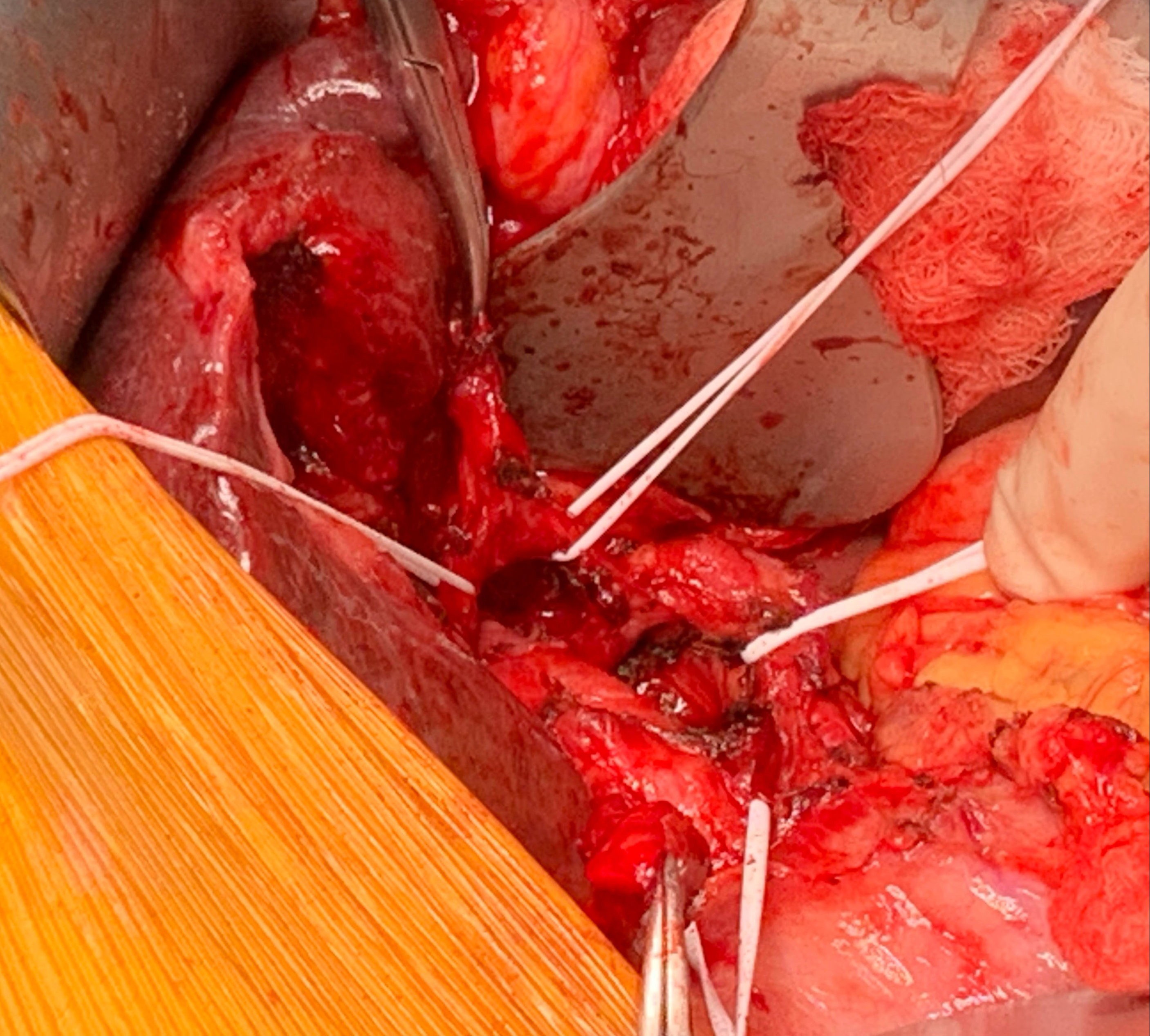
Figure: Relationship between the common hepatic duct (asterisk) and the mobilized anterior (solid
arrow) and posterior (dotted arrow) right hepatic artery branches.
arrow) and posterior (dotted arrow) right hepatic artery branches.
Disclosures:
David Iannitti indicated no relevant financial relationships.
Yifan Wang indicated no relevant financial relationships.
Erin Baker indicated no relevant financial relationships.
Matt Strand indicated no relevant financial relationships.
Stephen Deal indicated no relevant financial relationships.
David A. Iannitti, MD1, Yifan Wang, MD2, Erin Baker, MD1, Matt Strand, MD1, Stephen Deal, MD3. D0060 - Hepatolithiasis Caused by Right Hepatic Artery Branches Forming an Arterial Ring Compressing the Common Hepatic Duct, ACG 2022 Annual Scientific Meeting Abstracts. Charlotte, NC: American College of Gastroenterology.
