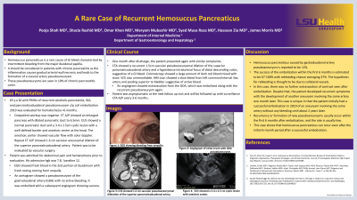
Back


Poster Session D - Tuesday Morning
Category: Biliary/Pancreas
D0084 - A Rare Case of Recurrent Hemosuccus Pancreaticus
Tuesday, October 25, 2022
10:00 AM – 12:00 PM ET
Location: Crown Ballroom

Has Audio

Pooja Shah, MD
Louisiana State University Health Sciences Center
Shreveport, LA
Presenting Author(s)
Pooja Shah, MD1, Shazia Rashid, MD2, Omar Khan, MD1, Maryam Mubashir, MD1, Syed Musa Raza, MD1, Hassaan Zia, MD3, James Morris, MD, FACG4
1Louisiana State University Health Sciences Center, Shreveport, LA; 2LSUHSC, Shreveport, LA; 3O-LSUS, Shreveport, LA; 4LSU Health Sciences Center, Shreveport, LA
Introduction: Hemosuccus pancreaticus is a rare cause of GI bleeds characterized by intermittent bleeding from the major duodenal papilla. Chronic pancreatitis, via arterial wall necrosis, can form a visceral pseudoaneurysm in 10% of cases. Here is a rare case of recurrent hemosuccus pancreaticus caused by a gastroduodenal artery (GDA) pseudoaneurysm, despite repeated embolization.
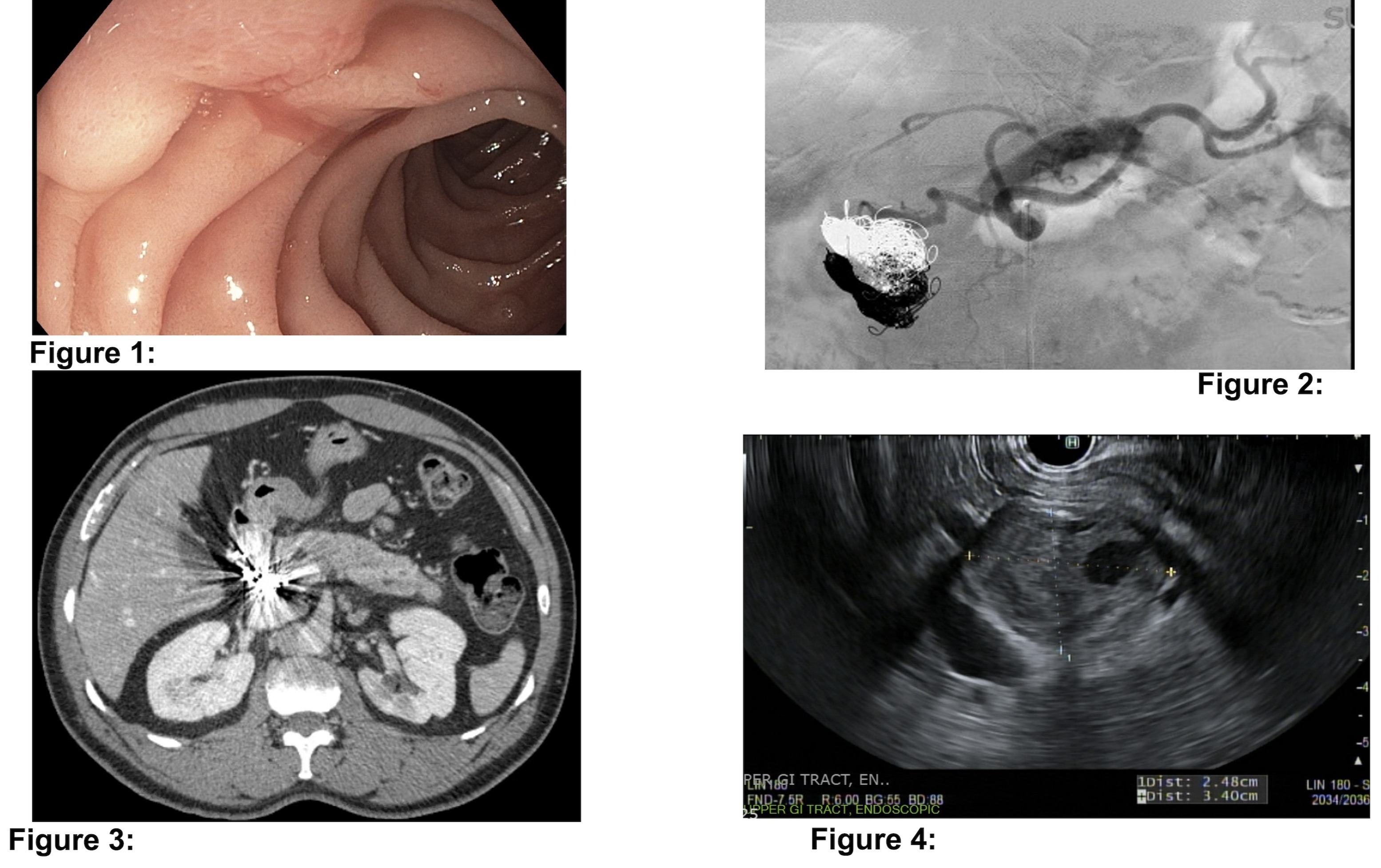
Case Description/Methods: A 65 y.o male with alcoholic pancreatitis and pancreaticoduodenal pseudoaneurysm status post embolization in 2019 was being evaluated for hematochezia for 6 months. The patient underwent several negative EGDs and colonoscopies. Video capsule endoscopy showed scant blood streaking in proximal small bowel. Initial CT showed an enlarged pancreas with dilated pancreatic duct. EUS showed a 3x2.5cm well-defined cystic lesion at the pancreatic head with an anechoic center with vascular flow. Repeat CT showed a 1.6cm saccular aneurysmal dilation of the superior pancreaticoduodenal artery (SPDA). Prior to follow-up, he was admitted with hematochezia, with his hemoglobin down from 12 to 7.8. EGD showed blood in the 2nd part of duodenum with fresh oozing from ampulla. An aortogram showed a non-bleeding pseudoaneurysm of the GDA, which was successfully embolized. One month later, he presented again with hematochezia. CTA showed a recurrent 1.5cm pseudoaneurysmal dilation of the SPDA and possible lower GI bleed. Colonoscopy showed dark red blood mixed with stool. NM scan showed a slow bleed from left common/internal iliac artery. An angiogram showed extravasation from the GDA, which was embolized along with the recurrent pseudoaneurysm. Seen 45 days after discharge, the patient was asymptomatic.
Discussion: Hemosuccus pancreaticus caused by GDA pseudoaneurysm is reported in less than 2% of population. Successful embolization in the first 6 months is estimated to be 67%, with rebleeding risk at 37%. Rebleeding is thought to be due to collateral vessels, but in this case, no extravasation was seen after each embolization. Our patient developed recurrent symptoms with development of an aneurysm involving the same artery 1 month later. Recurrence or formation of new pseudoaneurysms usually occur within the first 6 months after embolization. This case is unique in that the patient initially had a successful embolization in 2019 of an aneurysm involving the same artery without any bleeding for 2 years, showing that hemosuccus pancreaticus can recur even after the initial 6 month period after a successful embolization.

Disclosures:
Pooja Shah, MD1, Shazia Rashid, MD2, Omar Khan, MD1, Maryam Mubashir, MD1, Syed Musa Raza, MD1, Hassaan Zia, MD3, James Morris, MD, FACG4. D0084 - A Rare Case of Recurrent Hemosuccus Pancreaticus, ACG 2022 Annual Scientific Meeting Abstracts. Charlotte, NC: American College of Gastroenterology.
1Louisiana State University Health Sciences Center, Shreveport, LA; 2LSUHSC, Shreveport, LA; 3O-LSUS, Shreveport, LA; 4LSU Health Sciences Center, Shreveport, LA
Introduction: Hemosuccus pancreaticus is a rare cause of GI bleeds characterized by intermittent bleeding from the major duodenal papilla. Chronic pancreatitis, via arterial wall necrosis, can form a visceral pseudoaneurysm in 10% of cases. Here is a rare case of recurrent hemosuccus pancreaticus caused by a gastroduodenal artery (GDA) pseudoaneurysm, despite repeated embolization.
Case Description/Methods: A 65 y.o male with alcoholic pancreatitis and pancreaticoduodenal pseudoaneurysm status post embolization in 2019 was being evaluated for hematochezia for 6 months. The patient underwent several negative EGDs and colonoscopies. Video capsule endoscopy showed scant blood streaking in proximal small bowel. Initial CT showed an enlarged pancreas with dilated pancreatic duct. EUS showed a 3x2.5cm well-defined cystic lesion at the pancreatic head with an anechoic center with vascular flow. Repeat CT showed a 1.6cm saccular aneurysmal dilation of the superior pancreaticoduodenal artery (SPDA). Prior to follow-up, he was admitted with hematochezia, with his hemoglobin down from 12 to 7.8. EGD showed blood in the 2nd part of duodenum with fresh oozing from ampulla. An aortogram showed a non-bleeding pseudoaneurysm of the GDA, which was successfully embolized. One month later, he presented again with hematochezia. CTA showed a recurrent 1.5cm pseudoaneurysmal dilation of the SPDA and possible lower GI bleed. Colonoscopy showed dark red blood mixed with stool. NM scan showed a slow bleed from left common/internal iliac artery. An angiogram showed extravasation from the GDA, which was embolized along with the recurrent pseudoaneurysm. Seen 45 days after discharge, the patient was asymptomatic.
Discussion: Hemosuccus pancreaticus caused by GDA pseudoaneurysm is reported in less than 2% of population. Successful embolization in the first 6 months is estimated to be 67%, with rebleeding risk at 37%. Rebleeding is thought to be due to collateral vessels, but in this case, no extravasation was seen after each embolization. Our patient developed recurrent symptoms with development of an aneurysm involving the same artery 1 month later. Recurrence or formation of new pseudoaneurysms usually occur within the first 6 months after embolization. This case is unique in that the patient initially had a successful embolization in 2019 of an aneurysm involving the same artery without any bleeding for 2 years, showing that hemosuccus pancreaticus can recur even after the initial 6 month period after a successful embolization.

Figure: Figure 1: EGD showing bleeding from ampulla.
Figure 2: Angiogram of celiac trunk with GDA pseudoaneurysm.
Figure 3: CTA showed 1.5cm saccular pseudoaneurysmal dilatation of the superior pancreaticoduodenal artery.
Figure 4: EUS showed a 3.4 x 2.5 cm cystic lesion with anechoic center.
Figure 2: Angiogram of celiac trunk with GDA pseudoaneurysm.
Figure 3: CTA showed 1.5cm saccular pseudoaneurysmal dilatation of the superior pancreaticoduodenal artery.
Figure 4: EUS showed a 3.4 x 2.5 cm cystic lesion with anechoic center.
Disclosures:
Pooja Shah indicated no relevant financial relationships.
Shazia Rashid indicated no relevant financial relationships.
Omar Khan indicated no relevant financial relationships.
Maryam Mubashir indicated no relevant financial relationships.
Syed Musa Raza indicated no relevant financial relationships.
Hassaan Zia indicated no relevant financial relationships.
James Morris: Celgene – Grant/Research Support.
Pooja Shah, MD1, Shazia Rashid, MD2, Omar Khan, MD1, Maryam Mubashir, MD1, Syed Musa Raza, MD1, Hassaan Zia, MD3, James Morris, MD, FACG4. D0084 - A Rare Case of Recurrent Hemosuccus Pancreaticus, ACG 2022 Annual Scientific Meeting Abstracts. Charlotte, NC: American College of Gastroenterology.
