Back


Poster Session D - Tuesday Morning
Category: Colon
D0129 - Epiploic Appendagitis: A Red Herring?
Tuesday, October 25, 2022
10:00 AM – 12:00 PM ET
Location: Crown Ballroom

Has Audio

Anuroop Yekula, MD
Saint Vincent Hospital
Worcester, MA
Presenting Author(s)
Anuroop Yekula, MD1, Aakriti Soni, MD1, Mithil Gowda Suresh, MD2, Susan V. George, MD1
1Saint Vincent Hospital, Worcester, MA; 2St. Vincent Hospital, Worcester, MA
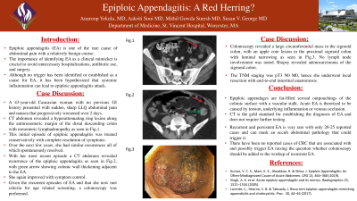
Introduction: Epiploic appendagitis (EA) is a rare cause of acute abdominal pain that has a relatively benign course. The importance of identifying EA as a clinical mimicker is crucial to avoid unnecessary hospitalizations, antibiotic use, and surgery. Although no trigger has been established as a cause for EA, it is hypothesized that systemic inflammation can lead to an EA attack.
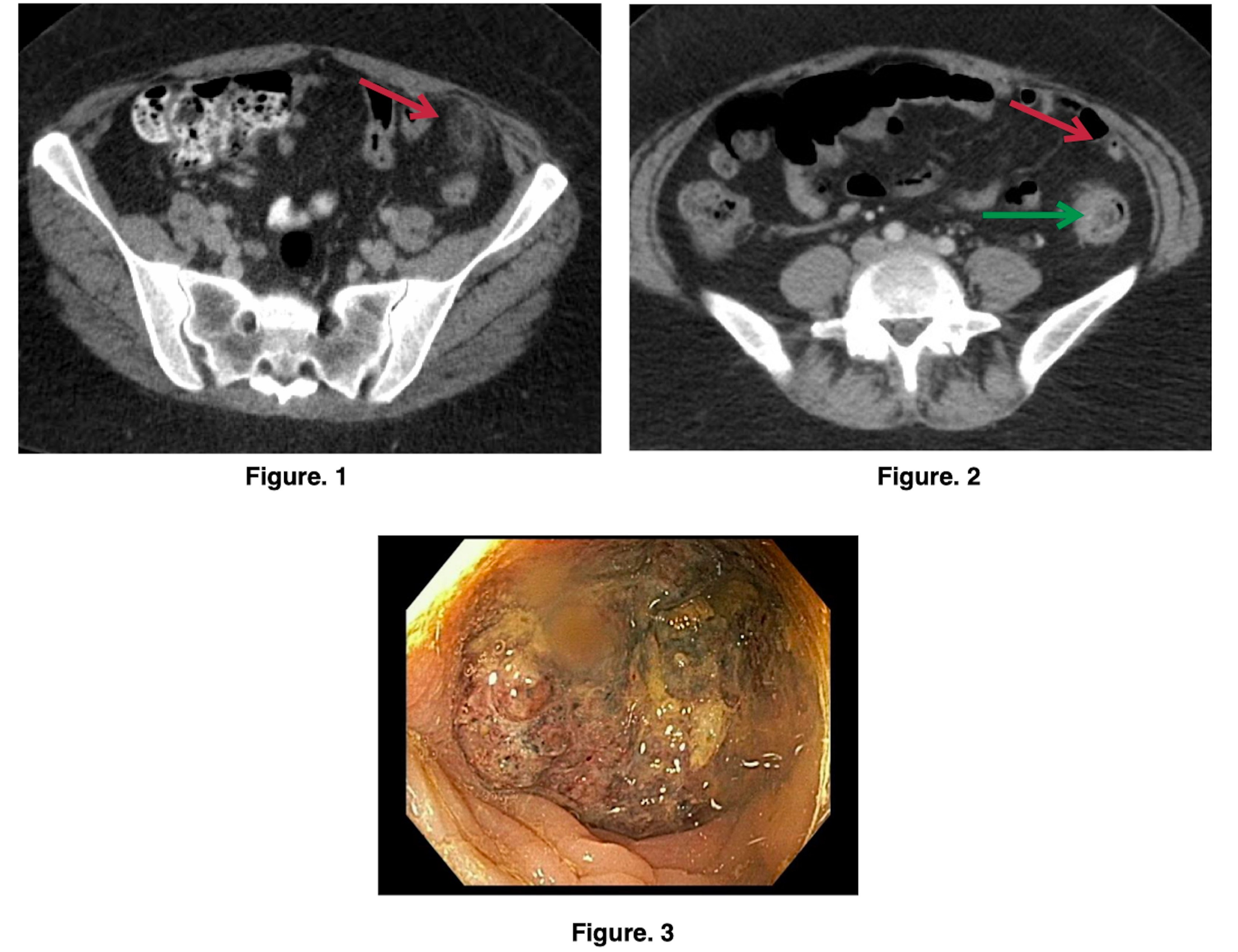
Case Description/Methods: A 43-year-old Caucasian female with no significant GI history presented with two days of sudden onset, sharp, non-radiating, worsening left lower quadrant (LLQ) with nausea. Initial blood work was unremarkable. CT abdomen revealed a hyper-attenuating ring lesion along the anti-mesenteric margin adjacent to the distal descending colon, along with mesenteric lymph nodes consistent with epiploic appendagitis. She was managed conservatively with complete resolution of symptoms.
A few years later, she presented again, with similar abdominal complaints. Repeat abdominal imaging showed recurrence of EA in the same location. Few shotty mesenteric lymph nodes were identified. She was treated conservatively for EA. A few more years passed, and she now had another episode of recurrent LLQ pain, CT abdomen showed findings consistent with EA along with a short segment of mural thickening and mild hyper-enhancement in the mid descending colon.
Colonoscopy revealed a large circumferential mass in the sigmoid colon with an apple core lesion in the proximal sigmoid colon with luminal narrowing. Biopsy revealed an adenocarcinoma. No lymph node involvement was noted. As the TNM staging was pT3 N0 M0, she underwent a sigmoidectomy with left colon and rectal end-to-end anastomosis.
Discussion: Epiploic appendages are fat-filled serosal outpouchings of the colonic surface. They are connected to the colon by a vascular stalk. Acute epiploic appendagitis is theorized to be caused by torsion, underlying inflammation, or venous occlusion of the appendage. CT scan is the gold standard for diagnosing EA and helps rule out other intra-abdominal pathologies.
Recurrent and persistent EA is very rare and may mask an underlying occult abdominal pathology. There have not been any reported cases of CRC that are associated with and possibly trigger EA. In patients with recurrent EA, after common causes of acute abdominal pain are ruled out, evaluation for intestinal/intraluminal pathologies, especially colorectal malignancy should be considered as they are not readily apparent on CT scans.

Disclosures:
Anuroop Yekula, MD1, Aakriti Soni, MD1, Mithil Gowda Suresh, MD2, Susan V. George, MD1. D0129 - Epiploic Appendagitis: A Red Herring?, ACG 2022 Annual Scientific Meeting Abstracts. Charlotte, NC: American College of Gastroenterology.
1Saint Vincent Hospital, Worcester, MA; 2St. Vincent Hospital, Worcester, MA
Introduction: Epiploic appendagitis (EA) is a rare cause of acute abdominal pain that has a relatively benign course. The importance of identifying EA as a clinical mimicker is crucial to avoid unnecessary hospitalizations, antibiotic use, and surgery. Although no trigger has been established as a cause for EA, it is hypothesized that systemic inflammation can lead to an EA attack.
Case Description/Methods: A 43-year-old Caucasian female with no significant GI history presented with two days of sudden onset, sharp, non-radiating, worsening left lower quadrant (LLQ) with nausea. Initial blood work was unremarkable. CT abdomen revealed a hyper-attenuating ring lesion along the anti-mesenteric margin adjacent to the distal descending colon, along with mesenteric lymph nodes consistent with epiploic appendagitis. She was managed conservatively with complete resolution of symptoms.
A few years later, she presented again, with similar abdominal complaints. Repeat abdominal imaging showed recurrence of EA in the same location. Few shotty mesenteric lymph nodes were identified. She was treated conservatively for EA. A few more years passed, and she now had another episode of recurrent LLQ pain, CT abdomen showed findings consistent with EA along with a short segment of mural thickening and mild hyper-enhancement in the mid descending colon.
Colonoscopy revealed a large circumferential mass in the sigmoid colon with an apple core lesion in the proximal sigmoid colon with luminal narrowing. Biopsy revealed an adenocarcinoma. No lymph node involvement was noted. As the TNM staging was pT3 N0 M0, she underwent a sigmoidectomy with left colon and rectal end-to-end anastomosis.
Discussion: Epiploic appendages are fat-filled serosal outpouchings of the colonic surface. They are connected to the colon by a vascular stalk. Acute epiploic appendagitis is theorized to be caused by torsion, underlying inflammation, or venous occlusion of the appendage. CT scan is the gold standard for diagnosing EA and helps rule out other intra-abdominal pathologies.
Recurrent and persistent EA is very rare and may mask an underlying occult abdominal pathology. There have not been any reported cases of CRC that are associated with and possibly trigger EA. In patients with recurrent EA, after common causes of acute abdominal pain are ruled out, evaluation for intestinal/intraluminal pathologies, especially colorectal malignancy should be considered as they are not readily apparent on CT scans.

Figure: Figure. 1: Red arrow showing an inflamed epiploic appendage during initial presentations.
Figure. 2: Red arrow showing epiploic appendagitis. Green arrow with colonic wall thickening in the descending colon, with adjacent EA.
Figure. 3: Colonoscopy showing the large friable mass in the sigmoid colon.
Figure. 2: Red arrow showing epiploic appendagitis. Green arrow with colonic wall thickening in the descending colon, with adjacent EA.
Figure. 3: Colonoscopy showing the large friable mass in the sigmoid colon.
Disclosures:
Anuroop Yekula indicated no relevant financial relationships.
Aakriti Soni indicated no relevant financial relationships.
Mithil Gowda Suresh indicated no relevant financial relationships.
Susan George indicated no relevant financial relationships.
Anuroop Yekula, MD1, Aakriti Soni, MD1, Mithil Gowda Suresh, MD2, Susan V. George, MD1. D0129 - Epiploic Appendagitis: A Red Herring?, ACG 2022 Annual Scientific Meeting Abstracts. Charlotte, NC: American College of Gastroenterology.

