Back


Poster Session D - Tuesday Morning
Category: Colon
D0139 - Empyema as a Rare Manifestation of Extraintestinal Clostridium difficile
Tuesday, October 25, 2022
10:00 AM – 12:00 PM ET
Location: Crown Ballroom

Has Audio

Ramzi Hassouneh, DO
Virginia Commonwealth University
Richmond, VA
Presenting Author(s)
Ramzi Hassouneh, DO1, Rachel Hart, DO2, Andrew Phillips, BS1, Shanedeep Gill, MD1, Sasha Mangray, MD1
1Virginia Commonwealth University, Richmond, VA; 2Vidant Medical Center, Greenville, NC
Introduction: Clostridium difficile is a gram-positive anaerobic bacteria that accounts for up to 25% of infectious diarrheal illnesses. Extra-intestinal C. difficile infection is exceedingly rare. Herein we report a case of C. difficile empyema.
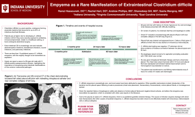
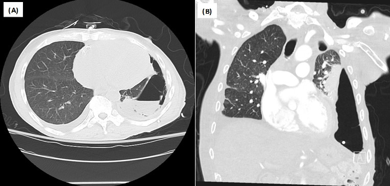
Case Description/Methods: A 69 year-old male with past history of chronic hypoxia (on 2L) and end-stage renal disease presented with difficulty breathing. He was tachypneic but oxygen saturation >90% on 2L. On review of systems, he endorsed 3-4 loose non-bloody bowel movements daily, dysphagia to solids and significant weight loss. Chest CT revealed a loculated large left pleural effusion with enhancement consistent with empyema and near complete collapse of the left lung (Figure). His pleural space was accessed and brown fluid was drained. Analysis of the pleural fluid showed pH 7.20, LDH 5745 U/L and protein 3.4 g/dL consistent with an exudative effusion. It was sent for culture and grew C. difficile at which time he was started on intravenous metronizadole. Blood cultures did not show any growth after 48 hours and C. difficile stool testing was negative. CT abdomen did not show evidence of fistulous connection between the pleural space and GI tract. His dysphagia raised concerns for aspiration prompting evaluation with an esophagram, which showed oropharyngeal dysphagia. He subsequently underwent EGD which was normal. He was given intrapleural fibrinolytic therapy and had a chest tube placed to assist with drainage of his empyema. The chest tube output improved within 72 hours and pleural fluid was reanalyzed and showed marked improvement (pH 7.51, LDH 74 U/L, total protein 1.7 g/dL) and no microrganism was detected on culture. The patient was transitioned to oral 500mg metronidazole every 8 hours for a total of 4 weeks and discharged.
Discussion: C. difficile empyema is exceedingly rare, and most cases have been attributed to aspiration. Other possible mechanisms include introduction into the pleural space following invasive procedures, enteropleural fistula, or hematogenous spread. Given the reported history of dysphagia to solids, we believe the empyema was transmitted via aspiration.
Due to the paucity of cases of C. difficile empyema, there is no published guideline directed therapy, however, it has been previously successfully treated with chest tube thoracostomy and metronidazole. While this infection is rare, it is important for clinicians to recognize and know how to treat.

Disclosures:
Ramzi Hassouneh, DO1, Rachel Hart, DO2, Andrew Phillips, BS1, Shanedeep Gill, MD1, Sasha Mangray, MD1. D0139 - Empyema as a Rare Manifestation of Extraintestinal Clostridium difficile, ACG 2022 Annual Scientific Meeting Abstracts. Charlotte, NC: American College of Gastroenterology.
1Virginia Commonwealth University, Richmond, VA; 2Vidant Medical Center, Greenville, NC
Introduction: Clostridium difficile is a gram-positive anaerobic bacteria that accounts for up to 25% of infectious diarrheal illnesses. Extra-intestinal C. difficile infection is exceedingly rare. Herein we report a case of C. difficile empyema.
Case Description/Methods: A 69 year-old male with past history of chronic hypoxia (on 2L) and end-stage renal disease presented with difficulty breathing. He was tachypneic but oxygen saturation >90% on 2L. On review of systems, he endorsed 3-4 loose non-bloody bowel movements daily, dysphagia to solids and significant weight loss. Chest CT revealed a loculated large left pleural effusion with enhancement consistent with empyema and near complete collapse of the left lung (Figure). His pleural space was accessed and brown fluid was drained. Analysis of the pleural fluid showed pH 7.20, LDH 5745 U/L and protein 3.4 g/dL consistent with an exudative effusion. It was sent for culture and grew C. difficile at which time he was started on intravenous metronizadole. Blood cultures did not show any growth after 48 hours and C. difficile stool testing was negative. CT abdomen did not show evidence of fistulous connection between the pleural space and GI tract. His dysphagia raised concerns for aspiration prompting evaluation with an esophagram, which showed oropharyngeal dysphagia. He subsequently underwent EGD which was normal. He was given intrapleural fibrinolytic therapy and had a chest tube placed to assist with drainage of his empyema. The chest tube output improved within 72 hours and pleural fluid was reanalyzed and showed marked improvement (pH 7.51, LDH 74 U/L, total protein 1.7 g/dL) and no microrganism was detected on culture. The patient was transitioned to oral 500mg metronidazole every 8 hours for a total of 4 weeks and discharged.
Discussion: C. difficile empyema is exceedingly rare, and most cases have been attributed to aspiration. Other possible mechanisms include introduction into the pleural space following invasive procedures, enteropleural fistula, or hematogenous spread. Given the reported history of dysphagia to solids, we believe the empyema was transmitted via aspiration.
Due to the paucity of cases of C. difficile empyema, there is no published guideline directed therapy, however, it has been previously successfully treated with chest tube thoracostomy and metronidazole. While this infection is rare, it is important for clinicians to recognize and know how to treat.

Figure: Figure. (A) Transverse CT chest demonstrating loculated left sided pleural effusion with indwelling intrapleural catheter. (B) Coronal CT chest demonstrating loculated left sided pleural effusion and near complete collapse of left lung.
Disclosures:
Ramzi Hassouneh indicated no relevant financial relationships.
Rachel Hart indicated no relevant financial relationships.
Andrew Phillips indicated no relevant financial relationships.
Shanedeep Gill indicated no relevant financial relationships.
Sasha Mangray indicated no relevant financial relationships.
Ramzi Hassouneh, DO1, Rachel Hart, DO2, Andrew Phillips, BS1, Shanedeep Gill, MD1, Sasha Mangray, MD1. D0139 - Empyema as a Rare Manifestation of Extraintestinal Clostridium difficile, ACG 2022 Annual Scientific Meeting Abstracts. Charlotte, NC: American College of Gastroenterology.
