Back


Poster Session D - Tuesday Morning
Category: Colon
D0151 - A Rare Case of Extraluminal Rectal Fecaloma
Tuesday, October 25, 2022
10:00 AM – 12:00 PM ET
Location: Crown Ballroom

Has Audio

Jelena Surla, MD
The Wright Center for Graduate Medical Education
Scranton, PA
Presenting Author(s)
Jelena Surla, MD1, Fouzia Oza, MD1, Ayla Benge, DO1, Mladen Jecmenica, MD2, Kristina Tanovic, MD1, Barry Pernikoff, MD3, Vikas Khurana, MD1, Aman Ali, MD1
1The Wright Center for Graduate Medical Education, Scranton, PA; 2Riverside Community Hospital, Riverside, CA; 3Commonwealth Health Physician Network, Wilkes-Barre, PA
Introduction: Fecaloma is usually an intraluminal stony mass of inspissated feces covered with layers of calcified shell. It can cause obstructive or compressive symptoms and sometimes even luminal rupture. It is usually managed conservatively and surgical removal is rarely needed. Most common sites for fecaloma are sigmoid colon and rectum. It is commonly associated with inflammatory bowel disease, Hirschsprung’s disease, Chagas disease, psychiatric and bedridden patients, neoplasm or idiopathic chronic constipation. We present a unique case of extraluminal fecalith presenting as subepithelial rectal mass.
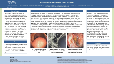
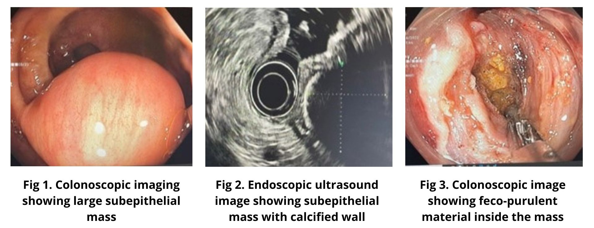
Case Description/Methods: A 60-year-old male with history of colon polyps underwent surveillance colonoscopy which showed a 3-4 cm firm rectal subepithelial mass that resembled a large lipoma or GIST (Fig. 1). Patient denied any symptoms including constipation or abdominal pain and had no family history of colon cancer. On endoscopic ultrasound (EUS) with radial and linear probe a subepithelial mass like lesion with calcified wall (Fig. 2) was noted. Multiple cold snare polypectomies were performed to de-roof the lesion as well as a deep "bite on bite/well biopsy technique" in order to get through the wall when feculent material and purulent debris was noted within the cavity (Fig. 3). Mucosal and muscular layer biopsies showed only vegetable fiber and fecal debris focally calcified consistent with fecalith. A CD 117 immunostain was negative excluding a GIST tumor. Historically this patient had an episode of complicated diverticulitis with phlegmon and microperforation in his 40s that was treated conservatively. Few months after that episode he had sigmoid colon resection with anastomosis in order to prevent future episodes.
Discussion: Although there are rare reported cases of extraluminal cecal fecalomas, to our knowledge this is the first reported case of extraluminal rectal fecaloma. We suspect that our patient developed fecal leakage through the suture line after sigmoid resection with anastomosis nearly 20 years ago. Over time, the leakage likely became a well-organized fecaloma with calcification. In summary, fecaloma is a differential diagnosis to consider when encountering a large subepithelial mass in the rectum.

Disclosures:
Jelena Surla, MD1, Fouzia Oza, MD1, Ayla Benge, DO1, Mladen Jecmenica, MD2, Kristina Tanovic, MD1, Barry Pernikoff, MD3, Vikas Khurana, MD1, Aman Ali, MD1. D0151 - A Rare Case of Extraluminal Rectal Fecaloma, ACG 2022 Annual Scientific Meeting Abstracts. Charlotte, NC: American College of Gastroenterology.
1The Wright Center for Graduate Medical Education, Scranton, PA; 2Riverside Community Hospital, Riverside, CA; 3Commonwealth Health Physician Network, Wilkes-Barre, PA
Introduction: Fecaloma is usually an intraluminal stony mass of inspissated feces covered with layers of calcified shell. It can cause obstructive or compressive symptoms and sometimes even luminal rupture. It is usually managed conservatively and surgical removal is rarely needed. Most common sites for fecaloma are sigmoid colon and rectum. It is commonly associated with inflammatory bowel disease, Hirschsprung’s disease, Chagas disease, psychiatric and bedridden patients, neoplasm or idiopathic chronic constipation. We present a unique case of extraluminal fecalith presenting as subepithelial rectal mass.
Case Description/Methods: A 60-year-old male with history of colon polyps underwent surveillance colonoscopy which showed a 3-4 cm firm rectal subepithelial mass that resembled a large lipoma or GIST (Fig. 1). Patient denied any symptoms including constipation or abdominal pain and had no family history of colon cancer. On endoscopic ultrasound (EUS) with radial and linear probe a subepithelial mass like lesion with calcified wall (Fig. 2) was noted. Multiple cold snare polypectomies were performed to de-roof the lesion as well as a deep "bite on bite/well biopsy technique" in order to get through the wall when feculent material and purulent debris was noted within the cavity (Fig. 3). Mucosal and muscular layer biopsies showed only vegetable fiber and fecal debris focally calcified consistent with fecalith. A CD 117 immunostain was negative excluding a GIST tumor. Historically this patient had an episode of complicated diverticulitis with phlegmon and microperforation in his 40s that was treated conservatively. Few months after that episode he had sigmoid colon resection with anastomosis in order to prevent future episodes.
Discussion: Although there are rare reported cases of extraluminal cecal fecalomas, to our knowledge this is the first reported case of extraluminal rectal fecaloma. We suspect that our patient developed fecal leakage through the suture line after sigmoid resection with anastomosis nearly 20 years ago. Over time, the leakage likely became a well-organized fecaloma with calcification. In summary, fecaloma is a differential diagnosis to consider when encountering a large subepithelial mass in the rectum.

Figure: Extraluminal Rectal Fecaloma
Disclosures:
Jelena Surla indicated no relevant financial relationships.
Fouzia Oza indicated no relevant financial relationships.
Ayla Benge indicated no relevant financial relationships.
Mladen Jecmenica indicated no relevant financial relationships.
Kristina Tanovic indicated no relevant financial relationships.
Barry Pernikoff indicated no relevant financial relationships.
Vikas Khurana: Aimloxy LLC – Owner/Ownership Interest.
Aman Ali indicated no relevant financial relationships.
Jelena Surla, MD1, Fouzia Oza, MD1, Ayla Benge, DO1, Mladen Jecmenica, MD2, Kristina Tanovic, MD1, Barry Pernikoff, MD3, Vikas Khurana, MD1, Aman Ali, MD1. D0151 - A Rare Case of Extraluminal Rectal Fecaloma, ACG 2022 Annual Scientific Meeting Abstracts. Charlotte, NC: American College of Gastroenterology.
