Back

Poster Session D - Tuesday Morning
Category: Colon
D0155 - Not an Ordinary Intussusception: A Sigmoid Diverticula With an Extra-Colonic Mass
Tuesday, October 25, 2022
10:00 AM – 12:00 PM ET
Location: Crown Ballroom


Shiva Poola, MD
ECU Health Medical Center/Brody School of Medicine
Greenville, NC
Presenting Author(s)
Shiva Poola, MD, Kara Regan, MD, Narasimha Swamy Gollol Raju, MD
ECU Health Medical Center/Brody School of Medicine, Greenville, NC
Introduction: Intussusception is defined as telescoping of a segment of the gastrointestinal tract into an adjacent lumen frequently found in childhood and are rarely found in adults. Etiologies of intussusception include benign or malignant tumors and idiopathic causes. Diverticula are rare findings for intussusception. This is a case of a patient found to have an intussusception due to a sigmoid diverticula and found to have a pelvic mass isolated outside of the diverticula.
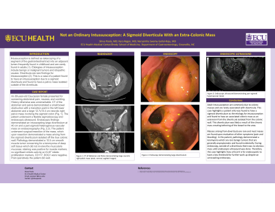
Case Description/Methods: An 80-year-old Caucasian female presented for worsening abdominal pain, nausea, and vomiting. History otherwise was unremarkable. CT of the abdomen and pelvis demonstrated a small bowel obstruction with a transition point in the left lower abdomen and a large 12.7x10.4 cm necrotic right pelvic mass involving the sigmoid colon (Fig. 1a). The patient underwent a flexible sigmoidoscopy and endoscopic ultrasound. Endoscopic findings demonstrated an intusscepting large diverticulum at 40 cm and a peri-sigmoid heterogenous vascular mass on endosonography (Fig. 1b/c). The patient underwent surgical resection of the mass, which upon resection demonstrated a mass arising from the sigmoid diverticulum isolated off the true colonic wall. Pathology demonstrated a 10.5 cm smooth muscle tumor concerning for a leiomyoma of deep soft tissue which did not involve the muscularis mucosae; staining was positive for muscle markers, no significant mitotic activity on Ki-67 mitotic proliferation markers, CD117, DOG1 were negative. Post operatively the patient did well.
Discussion: Adult Intussusception are commonly due to colonic masses and are rarely associated with diverticula. This case highlights a patient who was found to have a sigmoid diverticulum as the etiology for intussusception and found to have an associated colonic mass as an extension from the diverticula isolated from the colonic wall. This diverticulum was likely a result of the chronic mass creating tethering of the bowel to the area. Masses arising from diverticula are rare and the majority of mases are found upon evaluation of other symptoms (pain and bleeding). In this patient, pathology demonstrated a leiomyoma which are rare benign tumors that are generally asymptomatic and found incidentally. During endoscopy, outside of a diverticula there was no obvious mass until endoscopic ultrasound was done. Therefore, this case highlights how critical it is for endoscopists to have a low threshold for further work up despite an unrevealing endoscopy.

Disclosures:
Shiva Poola, MD, Kara Regan, MD, Narasimha Swamy Gollol Raju, MD. D0155 - Not an Ordinary Intussusception: A Sigmoid Diverticula With an Extra-Colonic Mass, ACG 2022 Annual Scientific Meeting Abstracts. Charlotte, NC: American College of Gastroenterology.
ECU Health Medical Center/Brody School of Medicine, Greenville, NC
Introduction: Intussusception is defined as telescoping of a segment of the gastrointestinal tract into an adjacent lumen frequently found in childhood and are rarely found in adults. Etiologies of intussusception include benign or malignant tumors and idiopathic causes. Diverticula are rare findings for intussusception. This is a case of a patient found to have an intussusception due to a sigmoid diverticula and found to have a pelvic mass isolated outside of the diverticula.
Case Description/Methods: An 80-year-old Caucasian female presented for worsening abdominal pain, nausea, and vomiting. History otherwise was unremarkable. CT of the abdomen and pelvis demonstrated a small bowel obstruction with a transition point in the left lower abdomen and a large 12.7x10.4 cm necrotic right pelvic mass involving the sigmoid colon (Fig. 1a). The patient underwent a flexible sigmoidoscopy and endoscopic ultrasound. Endoscopic findings demonstrated an intusscepting large diverticulum at 40 cm and a peri-sigmoid heterogenous vascular mass on endosonography (Fig. 1b/c). The patient underwent surgical resection of the mass, which upon resection demonstrated a mass arising from the sigmoid diverticulum isolated off the true colonic wall. Pathology demonstrated a 10.5 cm smooth muscle tumor concerning for a leiomyoma of deep soft tissue which did not involve the muscularis mucosae; staining was positive for muscle markers, no significant mitotic activity on Ki-67 mitotic proliferation markers, CD117, DOG1 were negative. Post operatively the patient did well.
Discussion: Adult Intussusception are commonly due to colonic masses and are rarely associated with diverticula. This case highlights a patient who was found to have a sigmoid diverticulum as the etiology for intussusception and found to have an associated colonic mass as an extension from the diverticula isolated from the colonic wall. This diverticulum was likely a result of the chronic mass creating tethering of the bowel to the area. Masses arising from diverticula are rare and the majority of mases are found upon evaluation of other symptoms (pain and bleeding). In this patient, pathology demonstrated a leiomyoma which are rare benign tumors that are generally asymptomatic and found incidentally. During endoscopy, outside of a diverticula there was no obvious mass until endoscopic ultrasound was done. Therefore, this case highlights how critical it is for endoscopists to have a low threshold for further work up despite an unrevealing endoscopy.

Figure: Figure 1: a (Top Left). CT Scan of abdomen and pelvis with large necrotic mass. b (Top Right): Endoscopic view of diverticula. c (Bottom Left): Endoscopic ultrasound of necrotic mass.
Disclosures:
Shiva Poola indicated no relevant financial relationships.
Kara Regan indicated no relevant financial relationships.
Narasimha Swamy Gollol Raju indicated no relevant financial relationships.
Shiva Poola, MD, Kara Regan, MD, Narasimha Swamy Gollol Raju, MD. D0155 - Not an Ordinary Intussusception: A Sigmoid Diverticula With an Extra-Colonic Mass, ACG 2022 Annual Scientific Meeting Abstracts. Charlotte, NC: American College of Gastroenterology.
