Back


Poster Session C - Monday Afternoon
Category: Esophagus
C0261 - Rare Case of 27-Year-Old With Esophageal Cancer
Monday, October 24, 2022
3:00 PM – 5:00 PM ET
Location: Crown Ballroom

Has Audio

Saakshi Joshi, MD
McLaren Macomb
Macomb, MI
Presenting Author(s)
Saakshi Joshi, MD, Alexandra Davies, DO, Johnathon Marcus, MD
McLaren Macomb, Macomb, MI
Introduction: Amongst esophageal cancers, neuroendocrine tumors represent 0.5 to 2.0% of cancers with a median age occurrence of 69 years old.¹ Our case represents a 27-year-old patient with neuroendocrine esophageal cancer.
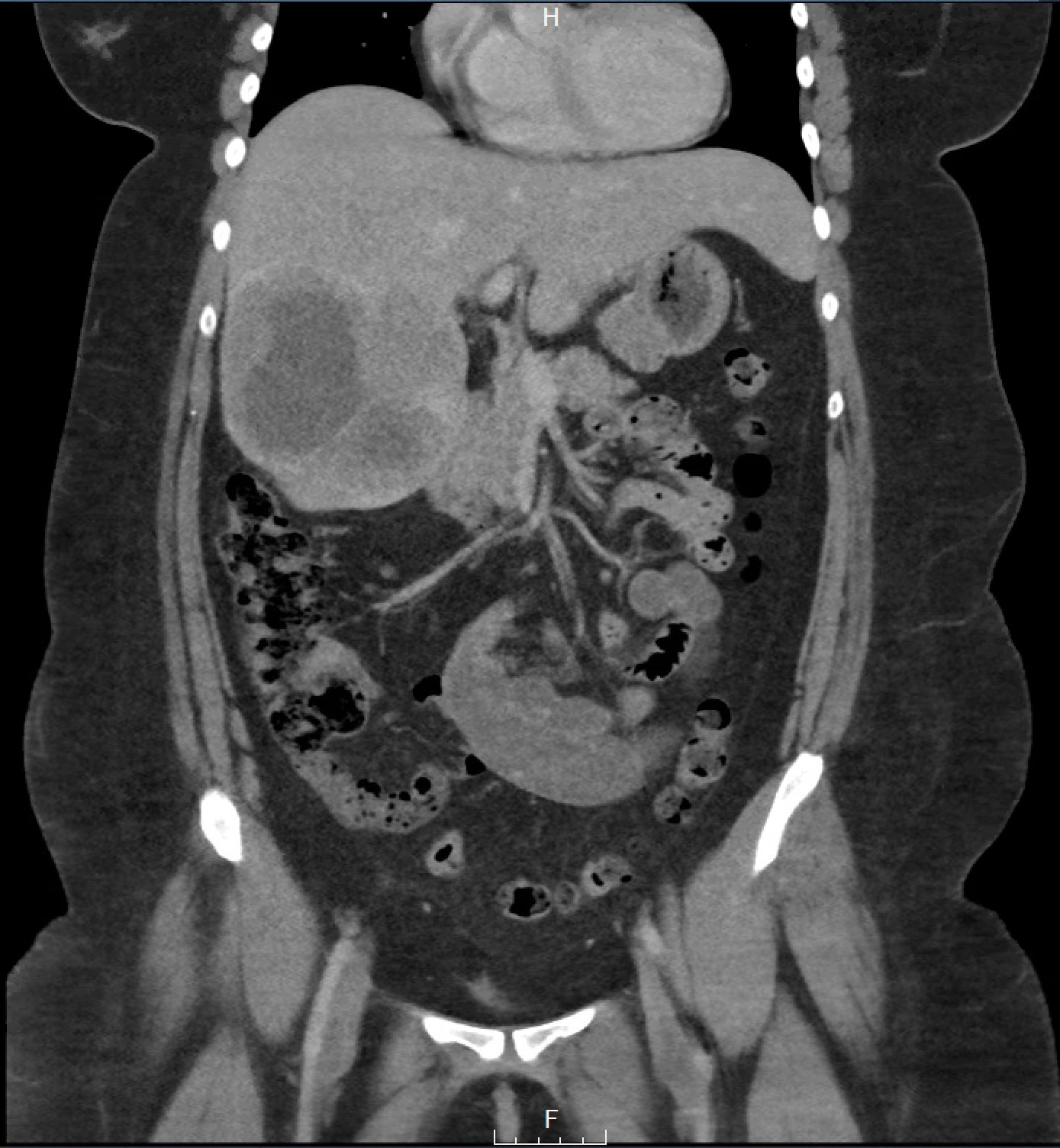
Case Description/Methods: 27 year old female with history of obesity presented to clinic for acid reflux with globus sensation and stated symptoms are worse after eating. The patient started on famotidine 20mg daily with no improvement, patient developed dysphagia to solids daily. Esophagogastroduodenoscopy (EGD) demonstrated a 4cm mass at distal esophagus bridging the gastroesophageal junction with a 2cm cratered ulceration at the distal end of the mass. Computed tomography of the abdomen showed abdominal adenopathy and 11cm metastatic lesion at the liver as seen in Figure 1. Pathology was chromogranin A positive representing a neuroendocrine tumor. Patient started on carboplatin and VP-16. Currently, the patient continuing to receive chemotherapy and will receive PET scan within the next month.
Discussion: As neuroendocrine tumors of the esophagus compromise a small portion of esophageal cancers with a female to male ratio of 1 to 3.7 emphasizing the importance of our case report.¹ Clinical manifestations include weight loss, anemia, and dysphagia. Endoscopic biopsy will confirm biopsy. Visualization on biopsy provides guidance on staging; early esophageal cancer presents as superficial plaques, nodules or ulcerations. Advanced lesions will appear as strictures, ulcerated masses, circumferential mass, or a large ulceration. Regardless of visualization on endoscopy, diagnosis is confirmed by biopsy. Biopsy confirms with an endocrine marker, example chromogranin A, synaptophysin, and CD 56. Staging then assists in guiding treatment. Endoscopic ultrasound is one of the most accurate techniques to assess with locoregional staging. Most common sites of metastasis are liver, lungs, bones and adrenal glands. There is controversy regarding the use of diagnostic laparoscopy but procedure generally reserved for patients who have potentially resectable tumor, T3 or T4 staging, Siewert II or III adenocarcinomas of the esophageal gastroduodenal junction, or when there is a suspicion for an intraperitoneal metastatic disease that cannot be confirmed otherwise.
[1] Egashira, Akinori et al. “Neuroendocrine carcinoma of the esophagus: Clinicopathological and immunohistochemical features of 14 cases.” PloS one vol. 12,3 e0173501. 13 Mar. 2017, doi:10.1371/journal.pone.0173501

Disclosures:
Saakshi Joshi, MD, Alexandra Davies, DO, Johnathon Marcus, MD. C0261 - Rare Case of 27-Year-Old With Esophageal Cancer, ACG 2022 Annual Scientific Meeting Abstracts. Charlotte, NC: American College of Gastroenterology.
McLaren Macomb, Macomb, MI
Introduction: Amongst esophageal cancers, neuroendocrine tumors represent 0.5 to 2.0% of cancers with a median age occurrence of 69 years old.¹ Our case represents a 27-year-old patient with neuroendocrine esophageal cancer.
Case Description/Methods: 27 year old female with history of obesity presented to clinic for acid reflux with globus sensation and stated symptoms are worse after eating. The patient started on famotidine 20mg daily with no improvement, patient developed dysphagia to solids daily. Esophagogastroduodenoscopy (EGD) demonstrated a 4cm mass at distal esophagus bridging the gastroesophageal junction with a 2cm cratered ulceration at the distal end of the mass. Computed tomography of the abdomen showed abdominal adenopathy and 11cm metastatic lesion at the liver as seen in Figure 1. Pathology was chromogranin A positive representing a neuroendocrine tumor. Patient started on carboplatin and VP-16. Currently, the patient continuing to receive chemotherapy and will receive PET scan within the next month.
Discussion: As neuroendocrine tumors of the esophagus compromise a small portion of esophageal cancers with a female to male ratio of 1 to 3.7 emphasizing the importance of our case report.¹ Clinical manifestations include weight loss, anemia, and dysphagia. Endoscopic biopsy will confirm biopsy. Visualization on biopsy provides guidance on staging; early esophageal cancer presents as superficial plaques, nodules or ulcerations. Advanced lesions will appear as strictures, ulcerated masses, circumferential mass, or a large ulceration. Regardless of visualization on endoscopy, diagnosis is confirmed by biopsy. Biopsy confirms with an endocrine marker, example chromogranin A, synaptophysin, and CD 56. Staging then assists in guiding treatment. Endoscopic ultrasound is one of the most accurate techniques to assess with locoregional staging. Most common sites of metastasis are liver, lungs, bones and adrenal glands. There is controversy regarding the use of diagnostic laparoscopy but procedure generally reserved for patients who have potentially resectable tumor, T3 or T4 staging, Siewert II or III adenocarcinomas of the esophageal gastroduodenal junction, or when there is a suspicion for an intraperitoneal metastatic disease that cannot be confirmed otherwise.
[1] Egashira, Akinori et al. “Neuroendocrine carcinoma of the esophagus: Clinicopathological and immunohistochemical features of 14 cases.” PloS one vol. 12,3 e0173501. 13 Mar. 2017, doi:10.1371/journal.pone.0173501

Figure: Figure 1: large heterogeneous lesion within the right hepatic lobe, measuring 10.5 x 11.5 cm which appears to demonstrate central necrosis
Disclosures:
Saakshi Joshi indicated no relevant financial relationships.
Alexandra Davies indicated no relevant financial relationships.
Johnathon Marcus indicated no relevant financial relationships.
Saakshi Joshi, MD, Alexandra Davies, DO, Johnathon Marcus, MD. C0261 - Rare Case of 27-Year-Old With Esophageal Cancer, ACG 2022 Annual Scientific Meeting Abstracts. Charlotte, NC: American College of Gastroenterology.
