Back


Poster Session D - Tuesday Morning
Category: General Endoscopy
D0283 - Deep Learning and Device-Assisted Enteroscopy: Automatic Detection of Pleomorphic Gastrointestinal Lesions in Device-Assisted Enteroscopy
Tuesday, October 25, 2022
10:00 AM – 12:00 PM ET
Location: Crown Ballroom

Has Audio

Miguel Mascarenhas, MD
Centro Hospitalar de S. João
Porto, Porto, Portugal
Presenting Author(s)
Miguel Mascarenhas, MD1, Joao Afonso, MD2, Tiago Ribeiro, MD1, Pedro Cardoso, MD1, Hélder Cardoso, MD1, João Ferreira, PhD3, Ana Andrade, MD1, Guilherme Macedo, MD, PhD1
1Centro Hospitalar de S. João, Porto, Porto, Portugal; 2Centro Hospitalar de São João, Porto, Porto, Portugal; 3FEUP: Faculdade de Engenharia da Universidade do Porto, Porto, Porto, Portugal
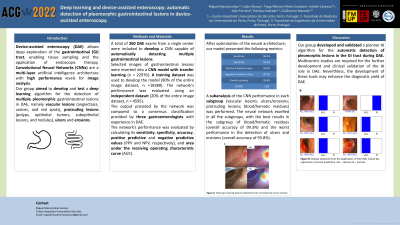
Introduction: Device-assisted enteroscopy (DAE) allows deep exploration of the gastrointestinal (GI) tract, enabling tissue sampling and the application of endoscopic therapy. Convolutional Neural Networks (CNNs) are a multi-layer artificial intelligence architecture with high performance levels for image analysis. Our group aimed to develop and test a deep learning algorithm for the detection of multiple pleomorphic gastrointestinal lesions in DAE, namely vascular lesions (angiectasia, varices, and red spots), protruding lesions (polyps, epithelial tumors, subepithelial lesions, and nodules), ulcers and erosions.
Methods: A total of 260 DAE exams from a single center were included to develop a CNN capable of automatically detecting multiple gastrointestinal lesions. Selected images of gastrointestinal lesions were inserted into a CNN model with transfer learning (n = 22976). A training dataset was used to develop the model (80% of the entire image dataset, n =18380). The network’s performance was evaluated using an independent dataset (20% of the entire image dataset, n = 4595). The output provided by the network was compared to a consensus classification provided by three gastroenterologists with experience in DAE. The network’s performance was evaluated by calculating its sensitivity, specificity, accuracy, positive predictive and negative predictive values (PPV and NPV, respectively), and area under the receiving operating characteristic curve (AUC).
Results: The trained CNN automatically detected gastrointestinal lesions with a sensitivity of 96.2%, a specificity of 95.0%, and a PPV and NPV of 95.6% and 95.7%. The overall accuracy was 95.6%. The AUC was 1.00. The CNN’s image processing time was 73 images per second. A subanalysis of the CNN performance in each subgroup (vascular lesions; ulcers/erosions; protruding lesions; blood/hematic residues) was performed. The neural network excelled in all the subgroups, with the best results in the subgroup of blood/hematic residues (overall accuracy of 99.3%) and the worst performance in the detection of ulcers and erosions (overall accuracy of 93.8%).
Discussion: Our group developed and validated a pionner AI algorithm for the automatic detection of pleomorphic lesions in the GI tract during DAE. Multicentric studies are required for the further development and clinical validation of the AI role in DAE. Neverthless, the development of these tools may enhance the diagnostic yield of DAE.

Disclosures:
Miguel Mascarenhas, MD1, Joao Afonso, MD2, Tiago Ribeiro, MD1, Pedro Cardoso, MD1, Hélder Cardoso, MD1, João Ferreira, PhD3, Ana Andrade, MD1, Guilherme Macedo, MD, PhD1. D0283 - Deep Learning and Device-Assisted Enteroscopy: Automatic Detection of Pleomorphic Gastrointestinal Lesions in Device-Assisted Enteroscopy, ACG 2022 Annual Scientific Meeting Abstracts. Charlotte, NC: American College of Gastroenterology.
1Centro Hospitalar de S. João, Porto, Porto, Portugal; 2Centro Hospitalar de São João, Porto, Porto, Portugal; 3FEUP: Faculdade de Engenharia da Universidade do Porto, Porto, Porto, Portugal
Introduction: Device-assisted enteroscopy (DAE) allows deep exploration of the gastrointestinal (GI) tract, enabling tissue sampling and the application of endoscopic therapy. Convolutional Neural Networks (CNNs) are a multi-layer artificial intelligence architecture with high performance levels for image analysis. Our group aimed to develop and test a deep learning algorithm for the detection of multiple pleomorphic gastrointestinal lesions in DAE, namely vascular lesions (angiectasia, varices, and red spots), protruding lesions (polyps, epithelial tumors, subepithelial lesions, and nodules), ulcers and erosions.
Methods: A total of 260 DAE exams from a single center were included to develop a CNN capable of automatically detecting multiple gastrointestinal lesions. Selected images of gastrointestinal lesions were inserted into a CNN model with transfer learning (n = 22976). A training dataset was used to develop the model (80% of the entire image dataset, n =18380). The network’s performance was evaluated using an independent dataset (20% of the entire image dataset, n = 4595). The output provided by the network was compared to a consensus classification provided by three gastroenterologists with experience in DAE. The network’s performance was evaluated by calculating its sensitivity, specificity, accuracy, positive predictive and negative predictive values (PPV and NPV, respectively), and area under the receiving operating characteristic curve (AUC).
Results: The trained CNN automatically detected gastrointestinal lesions with a sensitivity of 96.2%, a specificity of 95.0%, and a PPV and NPV of 95.6% and 95.7%. The overall accuracy was 95.6%. The AUC was 1.00. The CNN’s image processing time was 73 images per second. A subanalysis of the CNN performance in each subgroup (vascular lesions; ulcers/erosions; protruding lesions; blood/hematic residues) was performed. The neural network excelled in all the subgroups, with the best results in the subgroup of blood/hematic residues (overall accuracy of 99.3%) and the worst performance in the detection of ulcers and erosions (overall accuracy of 93.8%).
Discussion: Our group developed and validated a pionner AI algorithm for the automatic detection of pleomorphic lesions in the GI tract during DAE. Multicentric studies are required for the further development and clinical validation of the AI role in DAE. Neverthless, the development of these tools may enhance the diagnostic yield of DAE.

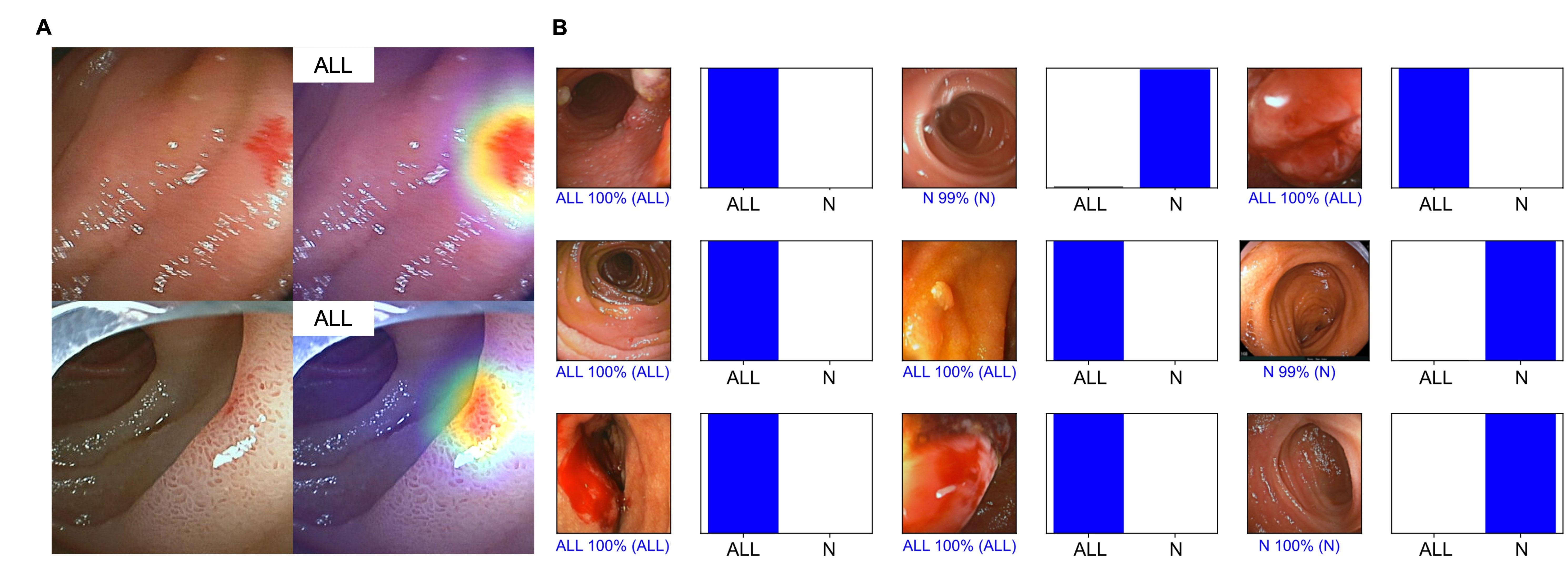
Figure: Figure 1: 1A – Heatmaps showing features detected by the convolutional neural network. 1B Output obtained from the application of the CNN. A blue bar represents a correct prediction. ALL – lesions; N – normal.
Disclosures:
Miguel Mascarenhas indicated no relevant financial relationships.
Joao Afonso indicated no relevant financial relationships.
Tiago Ribeiro indicated no relevant financial relationships.
Pedro Cardoso indicated no relevant financial relationships.
Hélder Cardoso indicated no relevant financial relationships.
João Ferreira indicated no relevant financial relationships.
Ana Andrade indicated no relevant financial relationships.
Guilherme Macedo indicated no relevant financial relationships.
Miguel Mascarenhas, MD1, Joao Afonso, MD2, Tiago Ribeiro, MD1, Pedro Cardoso, MD1, Hélder Cardoso, MD1, João Ferreira, PhD3, Ana Andrade, MD1, Guilherme Macedo, MD, PhD1. D0283 - Deep Learning and Device-Assisted Enteroscopy: Automatic Detection of Pleomorphic Gastrointestinal Lesions in Device-Assisted Enteroscopy, ACG 2022 Annual Scientific Meeting Abstracts. Charlotte, NC: American College of Gastroenterology.
